 Back
BackIntroduction to Microbiology: Core Concepts and Methods
Study Guide - Smart Notes
 Tailored notes based on your materials, expanded with key definitions, examples, and context.
Introduction to Microbiology
Definition and Scope
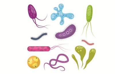
Microbiology is the scientific study of microorganisms, or microbes, which are typically too small to be seen with the naked eye. The field encompasses both cellular, living microorganisms (such as bacteria, archaea, fungi, protists, and helminths) and nonliving/noncellular entities (such as viruses and prions). Microbes inhabit nearly every environment on Earth, from deep-sea trenches to glaciers, and constitute at least half of Earth's biomass.
Microbe: A microscopic organism, which may be unicellular, multicellular, or acellular.
Pathogen: A microbe that causes disease.
Opportunistic pathogen: Causes disease only in a weakened host.
Applications: Microbiology is foundational in healthcare, agriculture, industry, and environmental sciences. Humans rely on microbes for food production, medication synthesis, and environmental remediation.
Cell Types in Microbiology
Prokaryotic vs. Eukaryotic Cells
Microorganisms are classified based on cell structure:
Prokaryotic cells: Lack a nucleus; include bacteria and archaea. Evolved ~3.5 billion years ago.
Eukaryotic cells: Have a nucleus; include fungi, protists, helminths, and all multicellular organisms.
Endosymbiotic theory: Explains the origin of eukaryotic organelles from ancestral prokaryotes.
Table: Living and Nonliving Agents Studied in Microbiology
Microbe | Cell Type | Notes |
|---|---|---|
Bacteria | Prokaryotic | Unicellular; pathogenic and nonpathogenic |
Archaea | Prokaryotic | Unicellular; nonpathogenic; extremophiles |
Protists | Eukaryotic | Unicellular/multicellular; pathogenic and nonpathogenic |
Fungi | Eukaryotic | Unicellular/multicellular; pathogenic and nonpathogenic |
Helminths | Eukaryotic | Multicellular; parasitic worms |
Viruses | Nonliving | DNA or RNA genome; infects cells |
Prions | Nonliving | Infectious proteins; cause neurodegenerative diseases |
Historical Foundations of Microbiology
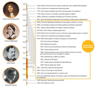
The Golden Age of Microbiology (1850–1920)
This era saw major advances in microscopy, microbial isolation, and disease causation. Key figures include Robert Hooke, Antonie van Leeuwenhoek, Louis Pasteur, and Robert Koch.
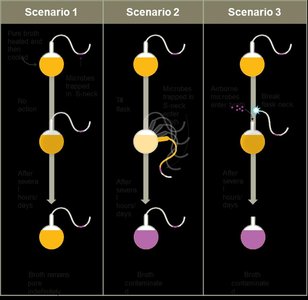
Spontaneous Generation vs. Biogenesis
Early scientists debated whether life arose spontaneously or from existing life. Experiments by Francesco Redi and Louis Pasteur disproved spontaneous generation, supporting biogenesis.
Spontaneous generation: Life arises from nonliving matter.
Biogenesis: Life arises from pre-existing life.
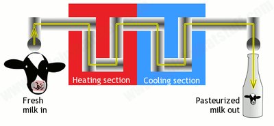
Pasteurization
Louis Pasteur developed pasteurization, a process of heating liquids to kill microbes and prevent spoilage.
Germ Theory of Disease and Koch’s Postulates
Germ Theory
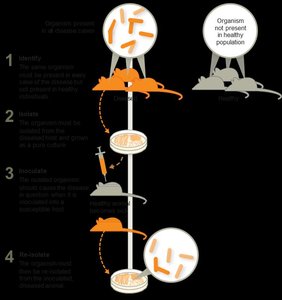
The germ theory of disease states that specific microbes cause specific diseases. Robert Koch established experimental criteria (Koch’s postulates) to link microbes to diseases.
Koch’s Postulates: Steps to prove a microbe causes a disease:
Microbe must be found in all cases of the disease.
Microbe must be isolated and grown in pure culture.
Pure culture must cause disease in a healthy host.
Microbe must be re-isolated from the experimentally infected host.
Aseptic Techniques and Hand Hygiene
Development of Aseptic Techniques
Medical professionals such as Ignaz Semmelweis, Joseph Lister, and Florence Nightingale pioneered aseptic techniques to prevent healthcare-associated infections (HAIs).
Hand washing
Wearing gloves
Sterilizing instruments
Decontaminating surfaces

The Scientific Method in Microbiology
Steps of the Scientific Method
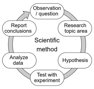
The scientific method is a systematic approach to investigating questions and testing hypotheses in microbiology.
Ask a question
Formulate a hypothesis
Collect and analyze data
Draw conclusions
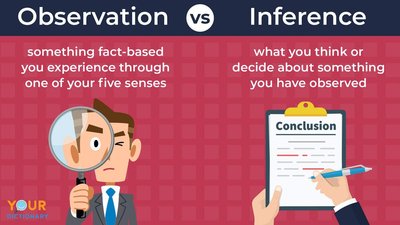
Observations vs. Conclusions
Observations are data collected through senses or instruments, while conclusions interpret these observations. Accurate conclusions require multiple observations.
Taxonomy and Classification
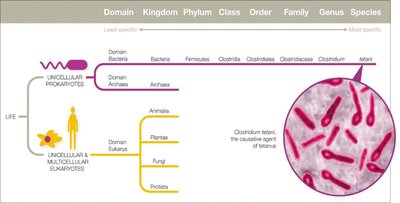
Taxonomic Hierarchy
Taxonomy is the science of classifying organisms. The hierarchy includes: Domain, Kingdom, Phylum, Class, Order, Family, Genus, Species.
Three domains: Bacteria, Archaea, Eukarya
Binomial nomenclature: Genus (capitalized) + species (lowercase), italicized (e.g., Escherichia coli)
Host–Microbe Interactions
Types of Symbiotic Relationships
Microbes and hosts interact in various ways:
Parasitism: Microbe harms the host.
Mutualism: Both benefit.
Commensalism: One benefits, the other is unaffected.
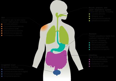
Normal microbiota (flora) are the collection of microbes living in and on the human body, often providing mutualistic benefits.
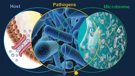
Microbiome and Human Health
The Human Microbiome Project (HMP) aims to characterize all microbes associated with humans. Normal microbiota help train the immune system, produce vitamins, aid digestion, and may influence mood and brain function.
Disruptions in Normal Microbiota
Antibiotic therapy can disrupt normal microbiota, allowing opportunistic pathogens to cause infections (e.g., Candida albicans yeast infections, antibiotic-associated diarrhea).
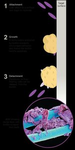
Biofilms
Formation and Importance
Biofilms are structured communities of microbes attached to surfaces and encased in a self-produced matrix. They are highly resistant to antibiotics and immune responses, and are implicated in 60–80% of human infections.
Develop on teeth, medical devices, water systems, etc.
Biofilm formation involves attachment, growth, and periodic release of planktonic cells.
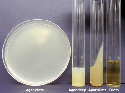
Microbial Culture and Growth Media
Growth Media Types
Growth media are nutrient mixtures used to cultivate microbes in the lab. Types include broths, agar plates, slants, and deeps. Agar is a common solidifying agent.
Aseptic Culture Techniques
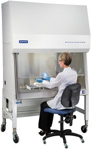
Aseptic techniques prevent contamination during microbial culture. This includes using sterile media, instruments, and protective clothing. Biological safety cabinets further reduce contamination risk.
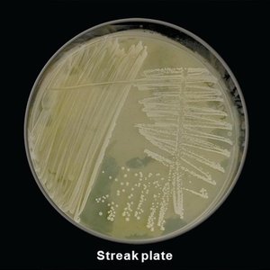
Streak Plate and Colony Isolation
The streak plate technique isolates individual colonies from a mixed sample, allowing for the study of pure cultures.
Microscopy and Staining Techniques
Staining Methods
Stains increase contrast in microscopic samples. Basic dyes (e.g., methylene blue, crystal violet) stain cells, while acidic dyes (e.g., nigrosin) stain backgrounds. Mordants (e.g., iodine) fix dyes to cells.
Types of Stains
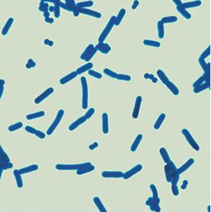
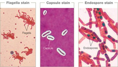
Simple stains: One dye; reveals cell shape and arrangement.
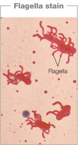
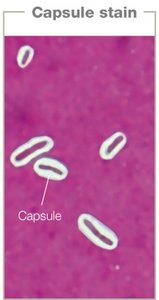
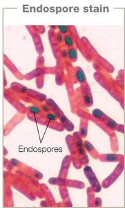
Structural stains: Highlight specific structures (flagella, capsules, endospores).
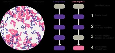
Differential stains: Distinguish between cell types (Gram stain, acid-fast stain).
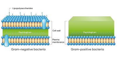
Gram Stain
The Gram stain differentiates bacteria based on cell wall structure:
Gram-positive: Thick peptidoglycan, no outer membrane; stains purple.
Gram-negative: Thin peptidoglycan, outer membrane; stains pink.
Acid-Fast Stain
Acid-fast staining identifies bacteria with waxy cell walls (e.g., Mycobacterium species). Acid-fast cells retain red dye after acid wash; non–acid-fast cells do not.
Microscopy Techniques
Light Microscopy
Light microscopes use visible light and lenses to magnify specimens. The compound light microscope is most common, with magnifications up to 1,500x and resolution of about 200 nm.
Types of Light Microscopy
Technique | Image Appearance | Notes |
|---|---|---|
Bright Field | Dark image on bright background | Requires staining or natural coloration |
Dark Field | Bright specimen on dark background | Visualizes unstained specimens |
Phase Contrast | Enhanced contrast, bright organelles | Visualizes live or dead, unstained specimens |
Differential Interference Contrast | 3D appearance | Uses polarized light |
Electron Microscopy
Electron microscopes use electron beams for much higher resolution (up to 0.2 nm). Two main types:
Transmission Electron Microscopy (TEM): 2D images of internal structures.
Scanning Electron Microscopy (SEM): 3D images of surfaces.
Fluorescence and Probe Microscopy
Fluorescence microscopy uses dyes that emit light under UV illumination. Immunofluorescence links dyes to antibodies for specific detection. Probe microscopy (e.g., atomic force) can visualize surfaces at the atomic level.
Clinical Application: Cholera Case Study
Summary
Cholera, caused by Vibrio cholerae, is a waterborne disease. Improved sanitation reduced cases by preventing water contamination. V. cholerae forms symbiotic relationships with copepods and can exist as part of the transient microbiota in humans. Rugose colony forms are more resistant to harsh conditions due to biofilm formation.
Key Terms and Concepts
Microbiome: The collective genomes of the microbes in a particular environment.
Symbiosis: Close association between different species.
Bioremediation: Use of microbes to clean up environmental contaminants.
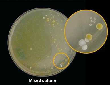
Pure culture: A culture containing only one species of microbe.
Colony: A visible mass of microbial cells derived from a single parent cell.
