 Back
BackIntroduction to Microbiology: Core Principles and Methods
Study Guide - Smart Notes
 Tailored notes based on your materials, expanded with key definitions, examples, and context.
Introduction to Microbiology
Definition and Scope
Microbiology is the scientific study of microorganisms, or microbes, which are typically too small to be seen with the naked eye. The field encompasses a wide range of organisms and entities, including cellular life forms and nonliving infectious agents.
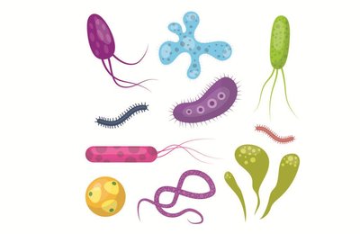
Microbes include bacteria, archaea, fungi, protists, helminths, viruses, and prions.
Microbes inhabit nearly every environment on Earth, from deep-sea trenches to glaciers.
Microbiology impacts healthcare, agriculture, industry, and environmental sciences.
Humans rely on microbes for food production, medication synthesis, and environmental remediation.
Types of Microbes
Microbe | Cell Type | Notes |
|---|---|---|
Bacteria | Prokaryotic | Unicellular; pathogenic and nonpathogenic |
Archaea | Prokaryotic | Unicellular; nonpathogenic; extremophiles |
Protists | Eukaryotic | Unicellular/multicellular; pathogenic and nonpathogenic |
Fungi | Eukaryotic | Unicellular/multicellular; pathogenic and nonpathogenic |
Helminths | Eukaryotic | Multicellular; parasitic worms |
Viruses | Non-cellular | Infect animal, plant, or bacterial cells; DNA or RNA genome |
Prions | Non-cellular | Infectious proteins; cause neurodegenerative diseases |
Historical Foundations of Microbiology
The Golden Age of Microbiology
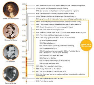
The period from 1850 to 1920 is known as the Golden Age of Microbiology, marked by major advances in microscopy, microbial isolation, and disease prevention.
Development of aseptic techniques and the germ theory of disease.
Key figures: Robert Hooke, Antonie van Leeuwenhoek, Louis Pasteur, Robert Koch, Florence Nightingale, Joseph Lister.
Spontaneous Generation vs. Biogenesis
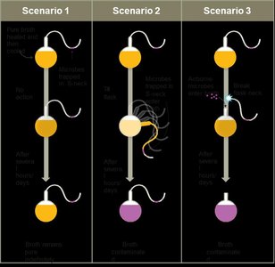
Early scientists debated whether life arose spontaneously or from existing life. Experiments by Redi and Pasteur provided evidence for biogenesis.
Spontaneous generation: Life arises from nonliving matter.
Biogenesis: Life arises from pre-existing life.
Pasteur's swan-neck flask experiment disproved spontaneous generation.
Pasteurization
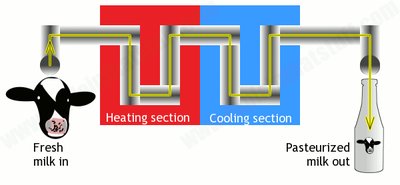
Louis Pasteur developed pasteurization, a process of heating liquids to reduce microbial load and prevent spoilage.
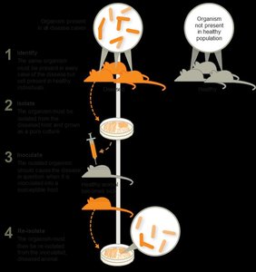
Germ Theory of Disease and Koch's Postulates
The germ theory states that specific microbes cause specific diseases. Robert Koch established a systematic method to link microbes to diseases.
Koch's postulates outline steps to prove a microbe causes a disease.
Not all microbes can be cultured, and new pathogens continue to emerge.
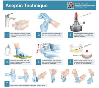
Aseptic Techniques and Infection Control
Development of Aseptic Techniques
Aseptic techniques are essential for preventing healthcare-associated infections (HAIs) and ensuring safe laboratory practices.
Hand washing, sterilizing instruments, and decontaminating surfaces are key practices.
Ignaz Semmelweis, Joseph Lister, and Florence Nightingale were pioneers in aseptic methods.
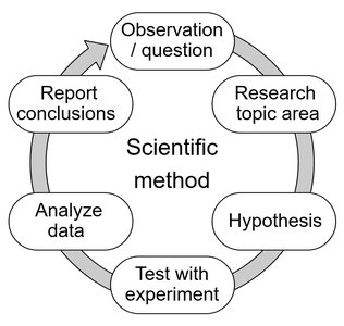
The Scientific Method in Microbiology
Principles and Application
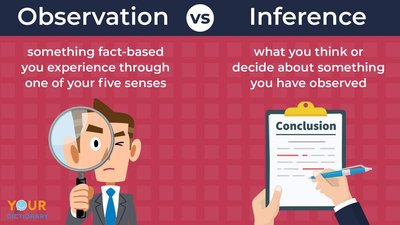
The scientific method is the foundation of microbiological research, involving observation, hypothesis formation, experimentation, and conclusion.
Observations are data collected via senses or instruments.
Conclusions interpret observations and must be based on sufficient evidence.
Distinguishing between observation and inference is critical in clinical settings.
Scientific Laws vs. Theories
Law: A precise statement or mathematical formula predicting a specific occurrence.
Theory: A hypothesis supported by extensive evidence explaining how and why phenomena occur.
Laws predict what happens; theories explain how and why.
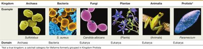
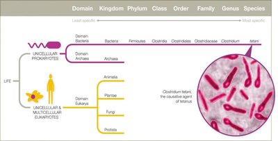
Taxonomy and Classification of Microbes
Taxonomic Hierarchy
Taxonomy organizes living organisms into hierarchical categories based on shared characteristics.
Hierarchy: Domain, Kingdom, Phylum, Class, Order, Family, Genus, Species.
Three domains: Bacteria, Archaea, Eukarya.
Classification systems have evolved from five to six kingdoms.
Scientific Nomenclature
Binomial nomenclature: Genus (capitalized) + species (lowercase), italicized (e.g., Escherichia coli).
Bergey’s Manual is a key reference for bacterial classification.
Strains are genetic variants within a species (e.g., E. coli K-12).
Host–Microbe Interactions
Symbiosis and Pathogenicity
Microbes interact with hosts in various ways, ranging from beneficial to harmful.
Parasitism: Microbe harms the host.
Mutualism: Both host and microbe benefit.
Commensalism: Microbe benefits; host is unaffected.
Pathogens are microbes that cause disease; most microbes are harmless or beneficial.
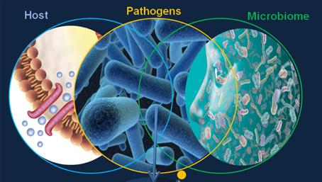
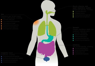
Normal Microbiota and the Human Microbiome
The normal microbiota (flora) consists of microbes that reside in and on the human body, playing essential roles in health and disease.
Functions: train immune system, produce vitamins, aid digestion, influence mood and brain function.
Microbiota composition varies by body site and individual.
Disruption (e.g., by antibiotics) can lead to infections by opportunistic pathogens.
Establishment and Disruption of Microbiota
Colonization begins at birth and is influenced by delivery mode and feeding.
Antibiotic therapy can disrupt normal microbiota, increasing infection risk (e.g., Candida albicans overgrowth).
Transient microbiota are temporary and removed by hygiene.
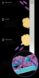
Biofilms
Formation and Significance
Biofilms are structured communities of microbes attached to surfaces and embedded in a self-produced matrix.
Biofilms can form on medical devices, teeth, water systems, and more.
They are highly resistant to antibiotics and immune responses.
Estimated 60–80% of human infections involve biofilms.
Microbial Culture and Laboratory Techniques
Growth Media
Growth media provide nutrients for microbial cultivation in the laboratory. Media types include broths, plates, slants, and deeps.

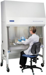
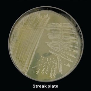
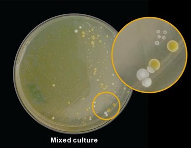
Aseptic Culture Techniques
Pure cultures are isolated using sterile techniques to prevent contamination.
Biological safety cabinets protect both samples and researchers.
Streak plate technique isolates individual colonies.
Microscopy and Staining Techniques
Staining Methods
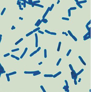
Simple stains: Use one dye to reveal cell shape and arrangement (e.g., methylene blue).
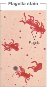
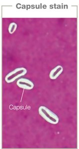
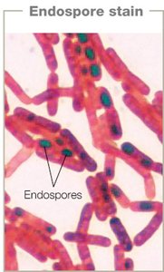
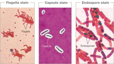
Structural stains: Highlight specific structures (e.g., flagella, capsules, endospores).
Differential stains: Distinguish between cell types (e.g., Gram stain, acid-fast stain).
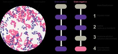
Gram Stain
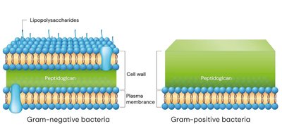
The Gram stain differentiates bacteria based on cell wall structure.
Gram-positive: thick peptidoglycan, no outer membrane, stains purple.
Gram-negative: thin peptidoglycan, outer membrane, stains pink.
Procedure: crystal violet, iodine, decolorizer, safranin.
Acid-Fast Stain
Distinguishes bacteria with waxy cell walls (e.g., Mycobacterium).
Acid-fast cells retain red dye after acid wash; non–acid-fast cells do not.
Ziehl-Neelsen method uses carbol-fuchsin, heat, acid-alcohol, and methylene blue.
Microscopy Techniques
Light Microscopy
Uses visible light and lenses to magnify specimens.
Types: bright field, dark field, phase contrast, differential interference contrast.
Resolution: ability to distinguish two points as separate; typical limit is 200 nm.
Oil immersion increases resolution at high magnification by reducing light scatter.
Comparison of Light Microscopy Techniques
Technique | Image | Notes |
|---|---|---|
Bright Field | Darker image on bright background | Requires staining or natural coloration |
Dark Field | Light sample on dark background | Visualizes unstained specimens |
Phase Contrast | Enhanced contrast, negative image | Visualizes live or dead specimens |
Differential Interference Contrast | 3D appearance | Uses polarized light |
Electron Microscopy
Uses electron beams for high-resolution imaging (up to 0.2 nm).
Transmission Electron Microscopy (TEM): 2D images of internal structures.
Scanning Electron Microscopy (SEM): 3D images of surfaces.
Specimens must be dead and often require special staining.
Fluorescence and Probe Microscopy
Fluorescence microscopy uses dyes that emit light under UV illumination.
Immunofluorescence links dyes to antibodies for specific detection.
Probe techniques (e.g., atomic force microscopy) visualize surfaces at the atomic level.
Clinical Application: Cholera Case Study
Summary and Key Concepts
Cholera is caused by Vibrio cholerae, a waterborne bacterium.
Proper sewage management reduces transmission by limiting water contamination.
Symbiotic relationships can be mutualistic (copepods and V. cholerae) or pathogenic (in humans).
Strain variation and environmental adaptation affect disease outcomes.
