Back
BackIntroduction to Microbiology: Foundations, Classification, and Laboratory Techniques
Study Guide - Smart Notes
 Tailored notes based on your materials, expanded with key definitions, examples, and context.
Introduction to Microbiology
Definition and Scope
Microbiology is the study of microorganisms (microbes), which are organisms too small to be seen with the naked eye. This field encompasses a wide range of life forms, including bacteria, archaea, fungi, protists, helminths, viruses, and prions. Microbiology is foundational to healthcare, agriculture, industry, and environmental sciences.
Microbes include both cellular (living) and noncellular (nonliving) entities.
Examples: Bacteria, Archaea, Fungi, Protists, Helminths, viruses, and prions.
Microbes inhabit nearly every environment on Earth, from deep-sea trenches to glaciers.
Importance of Microbes
Essential for food production, medication synthesis, and environmental remediation.
Most microbes are harmless or beneficial; less than 1% are pathogenic to humans.
A Brief History of Microbiology
Key Figures and Discoveries
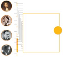
Robert Hooke: First to publish descriptions of cells (mid-1600s).
Antonie van Leeuwenhoek: Improved microscopes and first observed bacteria.
Louis Pasteur: Disproved spontaneous generation, developed pasteurization, and created vaccines for anthrax and rabies.
Robert Koch: Established the germ theory of disease and formulated Koch’s postulates.
Ignaz Semmelweis, Joseph Lister, Florence Nightingale: Pioneered aseptic techniques in healthcare.
Golden Age of Microbiology (1850–1920)
Development of advanced microscopes and laboratory techniques.
Discovery and characterization of many infectious agents.
Establishment of aseptic practices and immunizations.
Spontaneous Generation vs. Biogenesis
Historical Debate
Early scientists debated whether life arose spontaneously from nonliving matter (spontaneous generation) or from existing life (biogenesis).
Francesco Redi: Demonstrated that maggots on meat came from flies, not spontaneous generation.
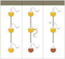
Louis Pasteur: Used S-necked flasks to show that microbes in the air, not spontaneous generation, caused contamination.
Germ Theory of Disease
Concept and Koch’s Postulates
The germ theory of disease states that specific microbes cause specific diseases. Robert Koch developed a systematic approach to identify the causative agents of infectious diseases.
Koch’s Postulates:
The same organism must be present in every case of the disease.
The organism must be isolated and grown in pure culture.
The isolated organism should cause the same disease in a healthy host.
The organism must be re-isolated from the experimentally infected host.
Aseptic Techniques and Hand Hygiene
Development and Importance
Ignaz Semmelweis: Advocated hand washing to prevent childbed fever.
Joseph Lister: Introduced sterilization of instruments and antiseptics in surgery.
Florence Nightingale: Established aseptic practices in nursing.
Aseptic techniques prevent healthcare-associated infections (HAIs) and include hand washing, wearing gloves, sterilizing instruments, and decontaminating surfaces.
The Scientific Method in Microbiology
Principles and Application
Begins with a testable question and hypothesis.
Data are collected through observation and experimentation.
Conclusions are drawn based on evidence.
Scientific law: Predicts what happens (often mathematical).
Scientific theory: Explains how and why phenomena occur, supported by extensive evidence.
Classifying Microbes and Their Interactions
Taxonomy and Nomenclature
Taxonomy is the science of classifying organisms based on shared characteristics. The taxonomic hierarchy includes:
Domain, Kingdom, Phylum, Class, Order, Family, Genus, Species
Binomial nomenclature assigns each organism a two-part scientific name: Genus (capitalized) and species (lowercase), both italicized (e.g., Escherichia coli).
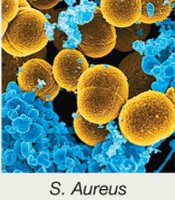
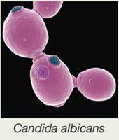

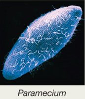
Strains and Species
Strain: Genetic variant of a species, often denoted by numbers/letters (e.g., E. coli K-12).
Prokaryotic species: Cells with ≥70% DNA similarity and ≥97% 16S rRNA sequence similarity.
Eukaryotic species: Organisms that can sexually reproduce together.
Symbiotic Relationships
Parasitism: Microbe harms the host.
Mutualism: Both microbe and host benefit.
Commensalism: Microbe benefits; host is unaffected.
Normal Microbiota and the Human Microbiome
Definition and Roles
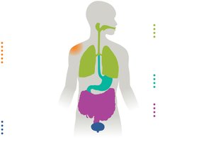
Normal microbiota (normal flora) are the microbes that reside in and on the human body, including bacteria, archaea, and eukaryotic microbes. They play crucial roles in health:
Train the immune system
Produce vitamins
Assist in digestion
Protect against pathogens by competitive exclusion
Establishment and Disruption
Colonization begins at birth and is influenced by delivery mode and feeding.
Antibiotic therapy can disrupt normal microbiota, leading to opportunistic infections (e.g., Candida albicans overgrowth).
Transient microbiota are temporary and removed by hygiene.
Biofilms
Formation and Clinical Importance
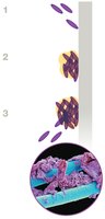
Biofilms are structured communities of microbes attached to surfaces and encased in a self-produced matrix. They are highly resistant to antibiotics and immune responses.
Form on teeth, medical devices, water systems, etc.
Responsible for 60–80% of human infections.
Laboratory Techniques: Growing, Staining, and Viewing Microbes
Culture Media
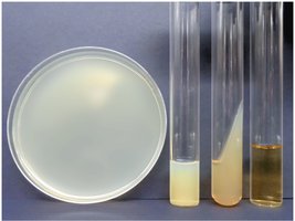
Growth media provide nutrients for microbial growth in the lab. Types include broths, agar plates, slants, and deeps.
Aseptic Culture Techniques
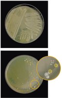
Essential for isolating pure cultures and preventing contamination.
Includes sterilizing media/instruments, decontaminating surfaces, and using protective gear.
Streak plate technique is used to isolate colonies.
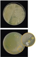
Staining Techniques
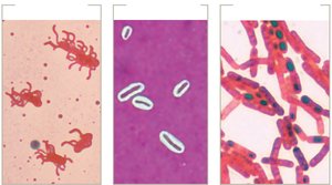
Simple stains: Use one dye to reveal cell shape, size, and arrangement.
Structural stains: Highlight specific structures (flagella, capsules, endospores).
Differential stains: Distinguish between cell types (e.g., Gram stain, acid-fast stain).
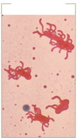
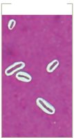
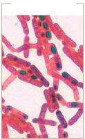
Gram Stain
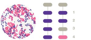
The Gram stain differentiates bacteria based on cell wall structure:
Gram-positive: Thick peptidoglycan, stains purple.
Gram-negative: Thin peptidoglycan, outer membrane, stains pink.
Acid-Fast Stain
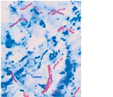
Distinguishes bacteria with waxy cell walls (e.g., Mycobacterium species) from those without.
Microscopy in Microbiology
Light Microscopy
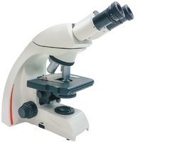
Uses visible light and lenses to magnify specimens. The compound light microscope is the most common type.
Resolution and Oil Immersion
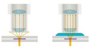
Resolution: Ability to distinguish two points as separate (limit ~200 nm for light microscopes).
Oil immersion: Increases resolution by reducing light refraction.
Types of Light Microscopy
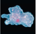
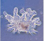
Bright field, dark field, phase contrast, and differential interference contrast.
Electron Microscopy
Transmission Electron Microscopy (TEM): Provides high-resolution 2D images of internal structures.
Scanning Electron Microscopy (SEM): Provides 3D images of specimen surfaces.
Fluorescence Microscopy
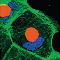
Uses fluorochromes to stain samples, which emit visible light under UV illumination.
Immunofluorescence uses antibodies linked to fluorescent dyes for specific detection.
Visual Summary