 Back
BackIntroduction to Microbiology: Foundations, Methods, and Microbial Interactions
Study Guide - Smart Notes
 Tailored notes based on your materials, expanded with key definitions, examples, and context.
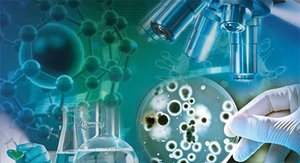
Introduction to Microbiology
Definition and Scope
Microbiology is the scientific study of microorganisms, or microbes, which are often invisible to the naked eye. The field encompasses both cellular, living microorganisms (such as bacteria, archaea, fungi, protists, and helminths) and nonliving/noncellular entities (such as viruses and prions). Some microorganisms, like certain fungi and helminths, are not always microscopic, but part of their life cycle is.
Microbes inhabit nearly every region of the planet, from deep-sea trenches to glaciers.
Microbiology spans health care, agriculture, industry, and environmental sciences.
Humans rely on microbes for food production, medication synthesis, and environmental remediation.
Fields of Microbiology
Bacteriology: Study of bacteria
Phycology: Study of algae
Mycology: Study of fungi
Protozoology: Study of protozoa
Parasitology: Study of parasites
Microbial genetics: Study of genetic information in microbes
Etiology: Study of disease causation
Microbial ecology: Study of microbe-environment relationships
Epidemiology: Study of disease frequency and distribution
Infection control: Study of nosocomial (hospital-acquired) infections
Chemotherapy: Research and use of drugs to treat diseases
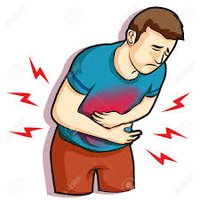
Microbes and Disease
Pathogens and Opportunistic Pathogens
Pathogens are microbes that cause disease. Of the thousands of microbial species, fewer than 1% are pathogenic to humans. Some pathogens always cause disease, while opportunistic pathogens only cause disease in weakened hosts.
History and Foundations of Microbiology
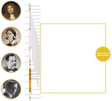
Golden Age of Microbiology (1850–1920)
This era saw major advances in microscopes, microbial isolation, and cultivation techniques. Key figures include Robert Hooke, Antonie van Leeuwenhoek, Florence Nightingale, Louis Pasteur, and Alexander Fleming.
1665: Robert Hooke observes eukaryotic cells
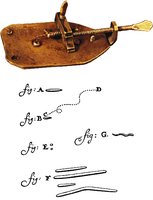
1670s: Antonie van Leeuwenhoek observes bacteria
1796: Edward Jenner vaccinates against smallpox
1860s: Florence Nightingale establishes aseptic practices
1864: Louis Pasteur disproves spontaneous generation
1876: Robert Koch proves a microbe causes disease (anthrax)
1928: Alexander Fleming discovers penicillin
Spontaneous Generation vs. Biogenesis
Early scientists debated whether life arose from nonliving matter (spontaneous generation) or from existing life (biogenesis). Louis Pasteur's experiments with S-necked flasks demonstrated that biogenesis is responsible for the propagation of life.
Germ Theory of Disease
Koch’s Postulates
The germ theory of disease states that microbes cause infectious diseases. Robert Koch developed a systematic approach to identify the causative agent of a disease, known as Koch’s postulates:
The same organism must be present in every case of the disease.
The organism must be isolated from the diseased host and grown as a pure culture.
The isolated organism should cause the disease when inoculated into a susceptible host.
The organism must be re-isolated from the inoculated, diseased animal.
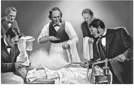
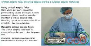
Aseptic Techniques and Hand Hygiene
Development of Aseptic Techniques
From the 1800s to 1900s, medical professionals emphasized aseptic techniques to prevent healthcare-acquired infections (HAIs). These include hand washing, wearing gloves, sterilizing instruments, and decontaminating surfaces.


Key Figures in Aseptic Practice
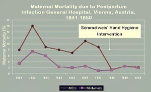
Ignaz Semmelweis: Advocated hand washing to reduce childbed fever mortality.
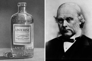
Joseph Lister: Developed aseptic surgery techniques, sterilizing instruments and wounds.
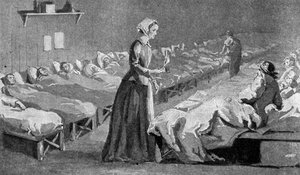
Florence Nightingale: Established aseptic techniques in nursing.
Scientific Method in Microbiology
Principles and Application
The scientific method guides investigations in microbiology. It involves forming a hypothesis, collecting and analyzing data, and drawing conclusions. Distinguishing between observations and conclusions is essential for accurate scientific assessment.
Law vs. Theory
Law: A precise statement or mathematical formula predicting a specific occurrence.
Theory: A hypothesis proven through repeated studies with consistent supporting conclusions.
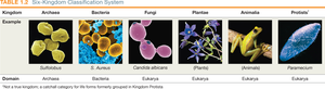
Classification of Microbes
Prokaryotic vs. Eukaryotic Cells
All life is cellular, classified as either prokaryotic or eukaryotic:
Prokaryotic organisms: Unicellular, lack a nucleus (bacteria and archaea).
Eukaryotic organisms: Unicellular or multicellular, have a distinct nucleus (animal, plant, fungal, protist).
Taxonomy and Binomial Nomenclature
Carl Linnaeus established the binomial nomenclature system: Genus (capitalized) and species (lowercase), both italicized (e.g., Escherichia coli).
Strain: Genetic variant of a species, often denoted by numbers/letters (e.g., E. coli K-12).
Taxonomic Hierarchy
Domain (broadest): Bacteria, Archaea, Eukarya
Kingdom: Animalia, Plantae, Fungi, Protista, Archaea, Bacteria
Host–Microbe Interactions
Symbiotic Relationships
Microbes and humans have evolved various symbiotic relationships:
Parasitism: Microbe harms the host
Mutualism: Both benefit
Commensalism: No perceived benefit or cost to the host
Normal Microbiota and Human Microbiome
The normal microbiota includes bacteria, archaea, and eukaryotic microbes residing in and on the human body. These microbes train the immune system, produce vitamins, aid digestion, and may influence mood and brain function.
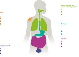
Microbiota varies by body region (skin, mouth, gut, genital/urinary tract).
Normal microbiota can include potential pathogens, but most are harmless and protect against disease by "crowding out" pathogens.
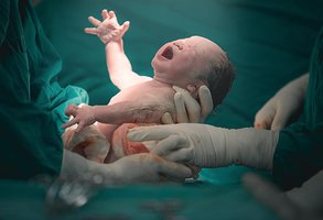
Establishing Normal Microbiota
Babies acquire microbiota during delivery and early interactions. Factors influencing microbiota development include delivery method and feeding type.

Disruptions in Normal Microbiota
Antibiotic therapy can disrupt normal microbiota, increasing susceptibility to opportunistic infections such as Candida albicans (yeast infection) and antibiotic-associated diarrhea.


Transient Microbiota
Transient microbiota are temporary microbes acquired from environmental contact and removed by hygiene practices.
Microbial Influence on Human Evolution
Host–microbe interactions have influenced human evolution. For example, carriers of the sickle cell gene have increased resistance to malaria, providing a survival advantage in endemic regions.
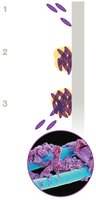
Biofilms
Formation and Characteristics
Biofilms are sticky communities of microbes attached to surfaces. They consist of single or diverse species and are difficult to penetrate due to their protective matrix.
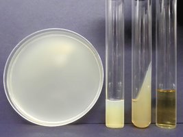
Microbial Cultivation and Growth Media
Growth Media Types
Growth media are nutrient mixtures supporting microbial growth in laboratory settings. Types include broths, plates, slants, and deeps. Agar is used as a solidifying agent for isolation.
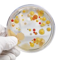
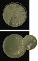
Aseptic Culture Techniques
Aseptic techniques are used to obtain pure cultures and prevent contamination. Colonies are groups of cells from a single parent cell; mixed cultures contain multiple colony types.

Staining and Microscopy
Staining Techniques
Stains increase contrast for microscopic observation. Staining methods include simple, structural, and differential stains.
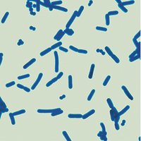
Simple Stains
Use one dye to determine cell size, shape, and arrangement.
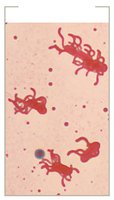
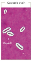
Structural Stains
Flagella stain: Reveals flagella arrangement
Capsule stain: Shows carbohydrate-based capsules as clear halos
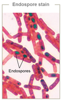
Endospore stain: Highlights dormant endospores in bacteria
Differential Stains
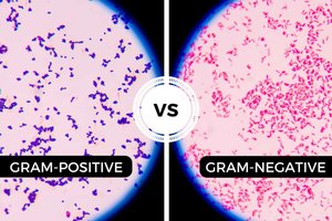
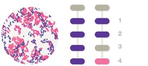
Gram stain: Classifies bacteria as Gram-positive (purple) or Gram-negative (pink)
Acid-fast stain: Distinguishes cells with waxy cell walls (e.g., Mycobacterium species)
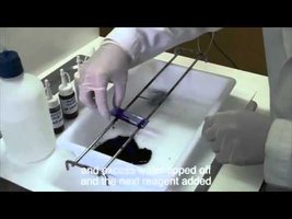
Gram Stain Procedure
Crystal violet (primary stain)
Iodine (mordant)
Acetone-alcohol (decolorizer)
Safranin (counterstain)
Microscopy
Light Microscopy
Uses visible light and lenses to magnify specimens. The compound light microscope is most common, with magnification up to 1,500× and resolution of about 200 nm.
Oil Immersion
Immersion oil is used with high-power objective lenses to reduce light refraction and improve image sharpness.
Types of Light Microscopy
Bright Field
Dark Field
Phase Contrast
Differential Interference Contrast
Electron Microscopy
Uses electron beams for high magnification and resolution. Two main types: transmission electron microscopes (TEM) and scanning electron microscopes (SEM).
Fluorescence Microscopy
Uses fluorescent dyes linked to antibodies for specific identification of microbes.
Visual Summary
Microbiology’s golden age established biogenesis and germ theory.
Classification systems organize microbes by shared features.
Symbiotic relationships include parasitism, mutualism, and commensalism.
Microscopy and staining techniques are central to microbial study.
Additional info: Some context and explanations were expanded for clarity and completeness, including definitions, examples, and academic background.
