 Back
BackIntroduction to Microbiology: Prokaryotes, Cell Structure, and Taxonomy
Study Guide - Smart Notes
 Tailored notes based on your materials, expanded with key definitions, examples, and context.
Introduction to Microbiology
Scope and Importance of Microbiology
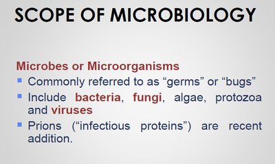
Microbiology is the study of microscopic organisms, including bacteria, fungi, algae, protozoa, and viruses. These organisms, often referred to as microbes or microorganisms, play crucial roles in the environment, industry, and health. The field also includes prions, which are infectious proteins.
Microbes are commonly called "germs" or "bugs" but include both beneficial and harmful species.
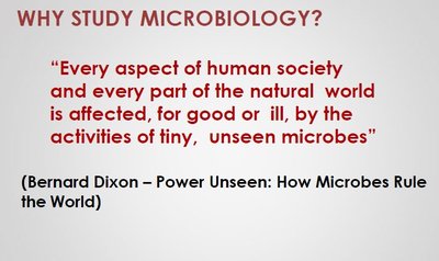
Microbiology impacts every aspect of human society and the natural world, influencing health, ecology, and biotechnology.
Quote: "Every aspect of human society and every part of the natural world is affected, for good or ill, by the activities of tiny, unseen microbes." (Bernard Dixon – Power Unseen: How Microbes Rule the World)
Size and Diversity of Microbes
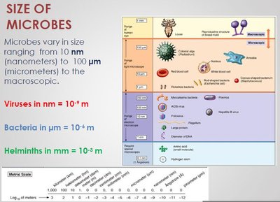
Microbes vary greatly in size, from nanometers (viruses) to millimeters (helminths). Understanding their scale is essential for appreciating their biological roles and laboratory handling.
Viruses: 10-9 meters (nanometers)
Bacteria: 10-6 meters (micrometers)
Helminths: 10-3 meters (millimeters)
Modern Uses of Microbes
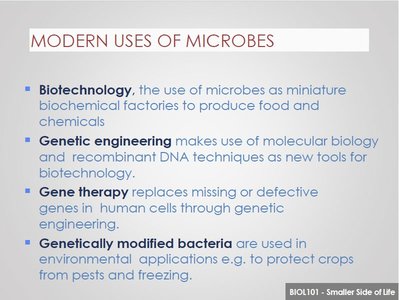
Microorganisms are utilized in various scientific and industrial applications:
Biotechnology: Microbes serve as biochemical factories for producing food and chemicals.
Genetic Engineering: Molecular biology and recombinant DNA techniques use microbes as tools for biotechnology.
Gene Therapy: Microbes are used to deliver or modify genes in human cells.
Genetically Modified Bacteria: Used in environmental applications, such as protecting crops from pests and freezing.
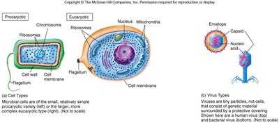
Prokaryotic and Eukaryotic Cell Structures
Comparative Cellular Structures
Microbial cells are classified as either prokaryotic or eukaryotic based on their structural complexity. Prokaryotes lack a membrane-bound nucleus and organelles, while eukaryotes possess these features.
Prokaryotes: Include bacteria and archaea; typically unicellular and smaller (0.5–5 µm).
Eukaryotes: Include fungi, algae, protozoa, and all multicellular organisms; larger (10–100 µm).

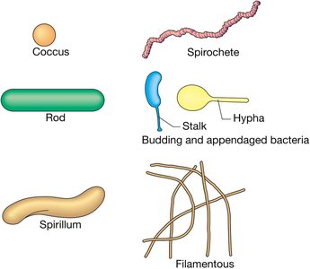
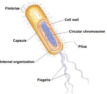
Prokaryotic Cell Morphology
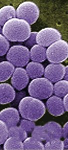
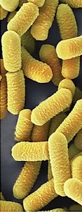
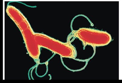
Prokaryotic cells exhibit various shapes and arrangements, which are important for identification and classification.
Cocci: Spherical; can occur singly (coccus), in pairs (diplococci), chains (streptococci), or clusters (staphylococci).
Bacilli: Rod-shaped; usually solitary or in chains (streptobacilli).
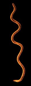
Spiral: Includes spirilla (rigid, spiral-shaped) and spirochetes (flexible, helical).
Microbial Taxonomy and Evolution
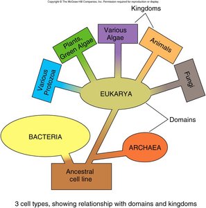
Three Domain System
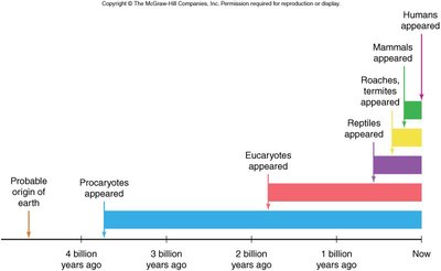
Modern taxonomy classifies all life into three domains based on genetic and biochemical characteristics: Bacteria, Archaea, and Eukarya. This system reflects evolutionary relationships and is supported by molecular data, such as ribosomal RNA sequences.
Bacteria: True bacteria, with peptidoglycan cell walls.
Archaea: Prokaryotes without peptidoglycan, often found in extreme environments.
Eukarya: All eukaryotic organisms, including plants, animals, fungi, and protists.
Taxonomic Hierarchy
Organisms are classified using a hierarchical system: Domain, Kingdom, Phylum, Class, Order, Family, Genus, Species. This system helps organize biological diversity and reflects evolutionary relationships.
Comparison of the Three Domains of Life
The following table summarizes key differences among Bacteria, Archaea, and Eukarya:
Characteristic | Bacteria | Archaea | Eukarya |
|---|---|---|---|
Nuclear envelope | Absent | Absent | Present |
Membrane-enclosed organelles | Absent | Absent | Present |
Peptidoglycan in cell wall | Present | Absent | Absent |
Membrane lipids | Unbranched hydrocarbons | Some branched hydrocarbons | Unbranched hydrocarbons |
RNA polymerase | One kind | Several kinds | Several kinds |
Initiator amino acid for protein synthesis | Formyl-methionine | Methionine | Methionine |
Introns in genes | Very rare | Present in some genes | Present in many genes |
Response to antibiotics | Growth inhibited | Not inhibited | Not inhibited |
Histones associated with DNA | Absent | Present in some species | Present |
Circular chromosome | Present | Present | Absent |
Growth at >100°C | No | Some species | No |
Prokaryotic Cell Walls and Gram Staining
Structure and Function of Prokaryotic Cell Walls
The cell wall is a critical structure in prokaryotes, providing shape, protection, and preventing lysis in hypotonic environments. The composition of the cell wall is a key feature for classification and antibiotic targeting.
Bacterial cell walls: Contain peptidoglycan (murein), a network of sugar polymers cross-linked by polypeptides. Main components are N-acetylglucosamine (NAG) and N-acetylmuramic acid (NAM).
Archaeal cell walls: Lack peptidoglycan; may contain glycoproteins, polysaccharides, or pseudomurein.
Eukaryotic cell walls: Made of cellulose (plants, algae) or chitin (fungi).
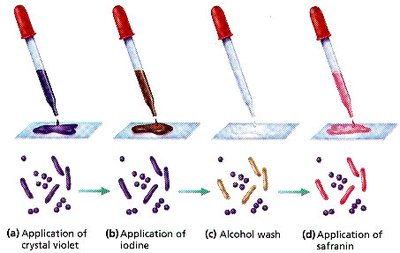
Gram Staining Procedure
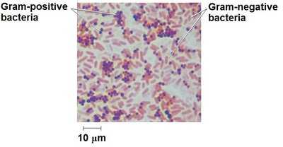
The Gram stain is a differential staining technique that classifies bacteria based on cell wall composition. Developed by Hans Christian Gram, it distinguishes between Gram-positive and Gram-negative bacteria.
Gram-positive bacteria: Thick peptidoglycan layer; stain purple.
Gram-negative bacteria: Thin peptidoglycan layer and an outer membrane containing lipopolysaccharides (LPS); stain pink/red.
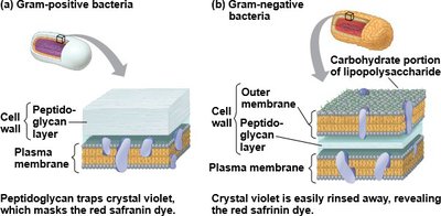
Significance of Gram-Negative Cell Wall
The outer membrane of Gram-negative bacteria contains lipopolysaccharides, which can be toxic and protect the bacteria from host defenses and certain antibiotics. Many antibiotics, such as penicillin, target peptidoglycan synthesis, making Gram-positive bacteria more susceptible. However, some Gram-positive bacteria have developed resistance to antibiotics.
Summary Table: Key Differences in Prokaryotic Cell Walls
Feature | Gram-Positive | Gram-Negative | Archaea |
|---|---|---|---|
Peptidoglycan | Thick layer | Thin layer | Absent |
Outer membrane | Absent | Present (LPS) | Absent |
Teichoic acids | Present | Absent | Absent |
Sensitivity to penicillin | High | Low | Variable |
Conclusion
Understanding the diversity, structure, and classification of microorganisms is foundational to microbiology. The differences in cell structure, especially cell wall composition, are critical for taxonomy, clinical diagnosis, and treatment strategies.
