 Back
BackIntroduction to Prokaryotic Cells – Structure, Classification, and Clinical Relevance
Study Guide - Smart Notes
 Tailored notes based on your materials, expanded with key definitions, examples, and context.
Introduction to Prokaryotic Cells
General Characteristics of Prokaryotes
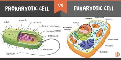
Prokaryotes are the smallest and most abundant organisms on Earth, characterized by their simple cellular organization. Unlike eukaryotes, prokaryotes lack a nucleus and membrane-bound organelles, with their DNA located in the cytoplasm.
Unicellular: All prokaryotes exist as single cells.
Genetic Material: DNA is found in the nucleoid region, not enclosed by a membrane.
Cell Size: Typically 0.5–2.0 µm in width, much smaller than eukaryotic cells (10–100 µm).
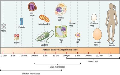
Additional info: The small size of prokaryotes is advantageous for nutrient uptake via diffusion, due to a high surface area-to-volume ratio.
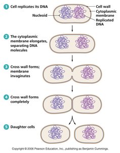
Prokaryote Reproduction: Binary Fission
Prokaryotes reproduce asexually through binary fission, a process that results in two genetically identical daughter cells.
Steps: DNA replication, elongation of the cell, formation of a septum, and division into two cells.
No genetic recombination: Offspring inherit genes from a single parent cell.
Doubling time: Varies with environmental conditions.
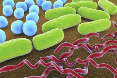
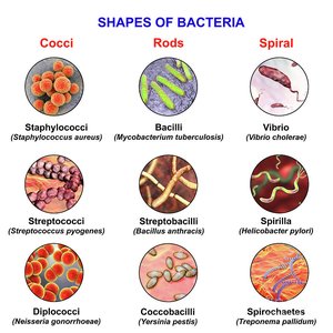
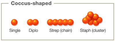
Prokaryote Morphology: Shapes and Arrangements
Prokaryotes exhibit diverse shapes and arrangements, which are often characteristic of specific genera or species and can aid in identification.
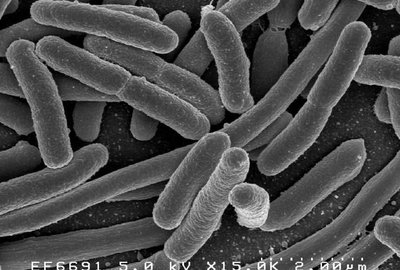
Common shapes: Cocci (spheres), bacilli (rods), spirilli (spirals).
Arrangements: Single, pairs (diplo-), chains (strepto-), clusters (staphylo-).
Monomorphic: Bacteria with a single, consistent shape.
Pleomorphic: Bacteria that can vary in shape, enhancing survival and transmission.

Classification of Prokaryotes
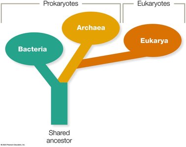
Domains of Life: Bacteria and Archaea
Prokaryotes are classified into two domains: Bacteria and Archaea. These domains are distinguished by genetic, biochemical, and structural differences.
Bacteria: Ubiquitous, diverse, and essential for life; most are non-pathogenic.
Archaea: Phenotypically similar to bacteria but genetically closer to eukaryotes; often extremophiles.
Eukarya: Branched off from Archaea ~2 billion years ago.
Archaea: Overview and Significance
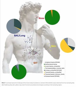
Archaea are found in a wide range of habitats, including extreme environments. They play critical roles in Earth's chemical cycles and are part of the normal microbiota of animals.
Extremophiles: Halophiles, thermophiles, psychrophiles, alkaliphiles, acidophiles.
Microbiota: Present in oral cavity, lung, skin, GI system; interactions are often mutual or commensal.
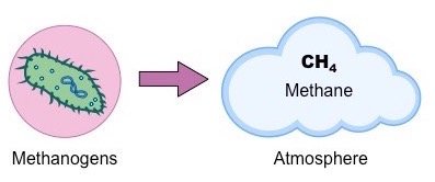
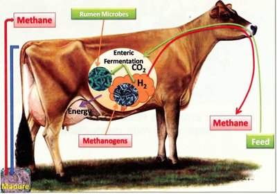
Methanogens: Aid digestion in ruminants and humans; important in bioremediation and waste treatment.

Bacteria: Overview
Bacteria are highly diverse unicellular organisms found in nearly every environment. Most are beneficial, with less than 5% being pathogenic.
Roles: Decomposition, bioremediation, food production, microbiome maintenance, and disease causation.
Example: Escherichia coli is a typical bacterium found in the human intestine.
Bacterial Structures
Internal Structures
Bacterial cells contain several key internal structures essential for their function and survival.
Cytoplasm: Gelatinous solution where cellular processes occur.
Nucleoid: Region containing the bacterial chromosome (usually circular DNA).
Ribosomes: 70S ribosomes for protein synthesis; smaller than eukaryotic 80S ribosomes.
Inclusion bodies: Storage sites for nutrients and sites of viral replication.
Cytoskeleton: Protein filaments providing structural support.
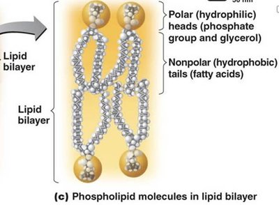
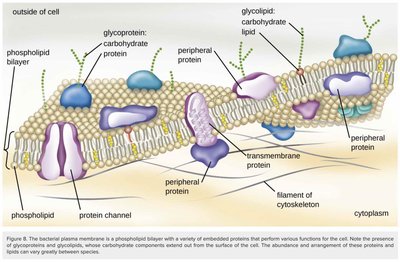
Plasma Membrane: Structure and Function
The plasma membrane is a flexible phospholipid bilayer that surrounds the cell, maintaining integrity and mediating interactions with the environment.
Fluid-mosaic model: Lipids and proteins move within the membrane, allowing dynamic responses.
Functions: Transport, anchoring, signaling, biosynthesis, and energy transduction.
Selective permeability: Allows passage of gases, water, and small molecules; larger molecules require transport proteins.
Transport Across the Plasma Membrane
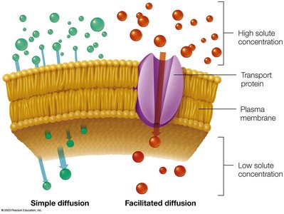
Bacteria use passive and active transport mechanisms to move substances across their plasma membrane.
Passive transport: No energy required; includes simple diffusion, facilitated diffusion, and osmosis.
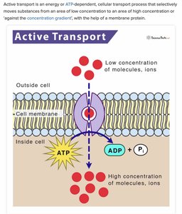
Active transport: Requires ATP; moves substances against their concentration gradient using carrier proteins.
External Structures
Structures outside the plasma membrane provide protection, aid in adhesion, and facilitate movement.
Cell wall: Rigid structure made of peptidoglycan; provides support and protection.
Glycocalyx: Capsule or slime layer; protects against desiccation and immune responses.
Fimbriae/Pili: Protein appendages for attachment, DNA transfer, and motility.
Flagella: Helical structures for motility; arrangements include monotrichous, lophotrichous, amphitrichous, peritrichous, and endoflagella.
Cell Wall Differences: Gram Stain
The Gram stain is a differential technique that classifies bacteria based on cell wall properties.
Gram-positive: Thick peptidoglycan layer, single membrane, teichoic acids.
Gram-negative: Thin peptidoglycan layer, outer membrane with lipopolysaccharide (LPS), porins.
Gram-indeterminate: Mycoplasmas (no cell wall), Mycobacteria (mycolic acid-rich wall).
Feature | Gram-positive | Gram-negative |
|---|---|---|
Peptidoglycan | Thick | Thin |
Outer Membrane | Absent | Present |
Teichoic Acids | Present | Absent |
LPS | Absent | Present |
Sensitivity to Penicillin | High | Low |
Glycocalyx: Capsule and Slime Layer
The glycocalyx is a carbohydrate-rich layer that aids in adhesion and protection. It exists as either a capsule (organized, dense) or slime layer (irregular, loose).
Capsule: Major virulence factor; protects against immune response.
Slime layer: Loosely attached; aids in surface attachment.
Filamentous Appendages: Fimbriae, Pili, and Flagella
These protein structures extend from the cell surface and are involved in attachment, motility, and genetic exchange.
Fimbriae/Pili: Short, hair-like; key for adherence and conjugation.
Flagella: Long, helical; provide motility via rotary movement.
Endospores
Some Gram-positive bacilli produce endospores, which are metabolically inactive, highly resistant structures that allow survival in harsh conditions.
Produced by: Bacillus, Clostridium, Clostridioides.
Clinical relevance: Endospores can persist in healthcare settings and cause disease.
Clinical Relevance and Review
Significance of Prokaryotes
Prokaryotes are essential for atmospheric oxygen production, decomposition, bioremediation, food production, microbiome maintenance, and disease causation.
Pathogenic bacteria: Cause infectious diseases; identification via Gram stain is crucial for diagnosis and treatment.
Summary Table: Key Differences Between Prokaryotic and Eukaryotic Cells
Feature | Prokaryotic Cell | Eukaryotic Cell |
|---|---|---|
Nucleus | Absent | Present |
Membrane-bound Organelles | Absent | Present |
Cell Size | 0.5–2.0 µm | 10–100 µm |
Cell Wall | Peptidoglycan (bacteria) | Cellulose (plants), chitin (fungi) |
Reproduction | Binary fission | Mitosis/meiosis |
Additional info: Use the provided learning objectives to guide your studying and complete relevant assignments for mastery of the material.
