 Back
BackIntroduction to Prokaryotic Cells: Structure, Function, and Clinical Relevance
Study Guide - Smart Notes
 Tailored notes based on your materials, expanded with key definitions, examples, and context.
Introduction to Prokaryotic Cells
Overview of Prokaryotes
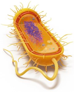
Prokaryotic cells are fundamental to the study of microbiology, representing the simplest and most ancient forms of life. Understanding their structure and function is essential for grasping microbial physiology, pathogenesis, and the basis for antibiotic action.
Prokaryotes include organisms in the domains Bacteria and Archaea.
They are unicellular, lack a membrane-bound nucleus, and do not possess membrane-bound organelles.
Prokaryotic cells are typically smaller than eukaryotic cells, which allows for efficient nutrient uptake and waste removal.
Domains of Life
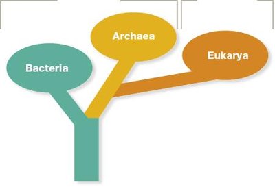
Life on Earth is classified into three domains: Bacteria, Archaea, and Eukarya. Prokaryotes are divided into Bacteria and Archaea, which share some similarities but also key differences.
Bacteria and Archaea are both prokaryotic but differ in cell wall composition, membrane lipids, and genetic machinery.
Eukarya includes all eukaryotic organisms, which have a true nucleus and organelles.
Comparison of Prokaryotic and Eukaryotic Cells
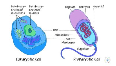
Understanding the differences between prokaryotic and eukaryotic cells is crucial for microbiology and medicine, especially in the context of antibiotic therapy.
Prokaryotes: Unicellular, lack nucleus, single circular chromosome, no organelles.
Eukaryotes: Uni- or multicellular, have nucleus, multiple linear chromosomes, possess organelles.
Both have DNA, ribosomes, cytoplasm, and a plasma membrane.
Prokaryotic Cell Size and Morphology
Cell Size and Surface Area-to-Volume Ratio
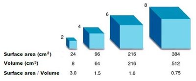
Prokaryotic cells are generally small, which maximizes their surface area-to-volume ratio, facilitating efficient nutrient diffusion and waste removal.
As cell size increases, the surface area-to-volume ratio decreases, limiting the rate of diffusion.
Small cells are more efficient at exchanging materials with their environment.

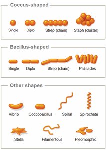
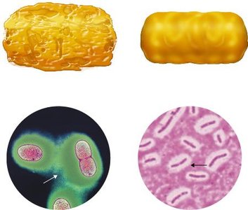
Shapes and Arrangements of Prokaryotes
Prokaryotes exhibit a variety of shapes (morphologies) and arrangements, which are important for identification and classification.
Bacilli: Rod-shaped
Cocci: Spherical
Vibrio: Comma-shaped
Stella: Star-shaped
Coccobacilli: Ovoid
Spirochetes: Spiral-shaped
Arrangements include diplococci (pairs), streptococci (chains), staphylococci (clusters), diplobacilli (pairs), streptobacilli (chains), and palisades (clusters).
Prokaryotic Cell Division
Binary Fission
Prokaryotic cells reproduce asexually by binary fission, resulting in two genetically identical daughter cells.
Steps of binary fission:
DNA is replicated.
Cell grows and elongates.
A septum forms at the midpoint.
The cell is partitioned into two.
Two daughter cells are separated.
This process is asexual and does not involve spore formation for reproduction.
Prokaryotic Cell Envelope and Transport Mechanisms
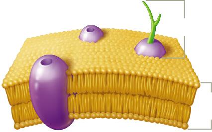
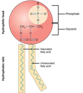
Plasma Membrane Structure and Function
The plasma membrane is a phospholipid bilayer with embedded proteins, providing selective permeability and structural integrity.
Hydrophilic heads face outward; hydrophobic tails face inward.
Proteins serve as transporters, anchors, receptors, and enzymes.
Small, non-charged molecules diffuse freely; ions and large molecules require transport proteins.


Cell Wall Structure and Differences
The cell wall provides rigidity and protection. Its composition differs between Bacteria and Archaea.
Bacteria: Cell wall contains peptidoglycan (linear fatty acids).
Archaea: Cell wall contains pseudopeptidoglycan (branched fatty acids); some form lipid monolayers for extreme environments.
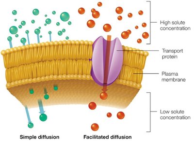
Transport Across the Cell Envelope
Prokaryotic cells use passive and active transport mechanisms to move substances across their membranes.
Passive transport: No energy required; includes simple diffusion, facilitated diffusion, and osmosis.
Active transport: Requires energy; moves substances against concentration gradients and includes primary/secondary active transport and group translocation.
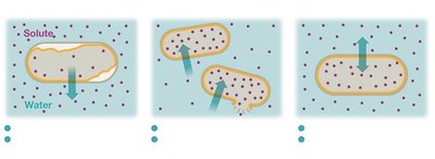
Osmosis and Tonicity
Osmosis is the movement of water across a semipermeable membrane, influenced by the solute concentration of the environment.
Hypertonic: Water leaves the cell, causing plasmolysis.
Hypotonic: Water enters the cell, possibly causing lysis if the cell wall is damaged.
Isotonic: No net water movement; cell remains stable.
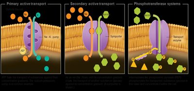
Active Transport Mechanisms
Active transport moves substances against their concentration gradients using energy, often via specialized proteins.
Includes primary active transport (e.g., Na+/K+ pump), secondary active transport (symporters/antiporters), and group translocation (phosphotransferase systems).
External Structures of Prokaryotic Cells
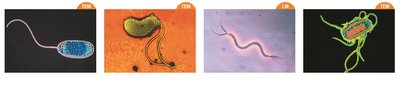
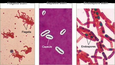
Flagella
Flagella are long, whip-like structures used for motility. They are composed of flagellin and can be arranged in various patterns.
Monotrichous: Single flagellum
Lophotrichous: Cluster at one pole
Amphitrichous: One or more at both poles
Peritrichous: Distributed over the entire cell surface
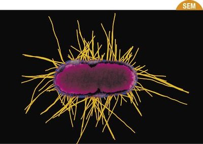
Fimbriae and Pili
Fimbriae are short, bristle-like structures that aid in adhesion and biofilm formation. Pili are longer, less numerous, and involved in adhesion, movement, and gene transfer (conjugation).

Glycocalyx
The glycocalyx is a carbohydrate-rich layer outside the cell wall, which can be a loosely associated slime layer or a well-organized capsule. It aids in adhesion, protection from desiccation, and resistance to antibiotics.
Staining Techniques and Cell Wall Differences
Simple and Differential Staining
Staining enhances visualization of cells under the microscope. Differential stains, such as the Gram stain, distinguish between cell wall types.
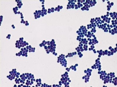
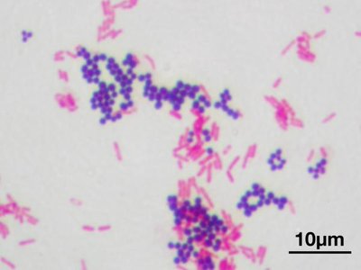
Gram Stain Procedure and Cell Wall Structure
The Gram stain differentiates bacteria based on cell wall structure:
Gram-positive: Thick peptidoglycan layer, no outer membrane, stains purple.
Gram-negative: Thin peptidoglycan layer, outer membrane with lipopolysaccharides (LPS), stains pink/red.
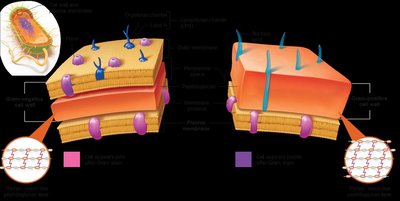
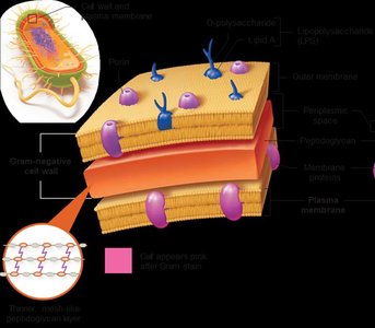
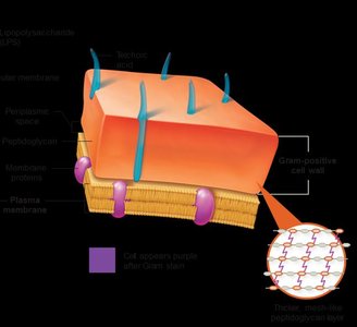
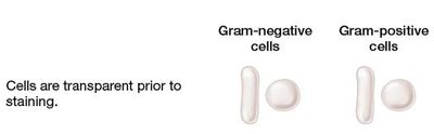

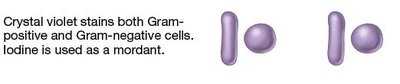
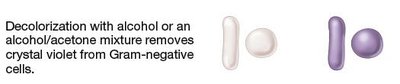
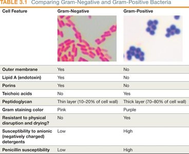
Table: Comparison of Gram-Negative and Gram-Positive Bacteria
Cell Feature | Gram-Negative | Gram-Positive |
|---|---|---|
Outer membrane | Yes | No |
Lipid A (endotoxin) | Yes | No |
Porins | Yes | No |
Teichoic acids | No | Yes |
Peptidoglycan | Thin layer (10–20%) | Thick layer (70–80%) |
Gram staining color | Pink | Purple |
Physical resistance | No | Yes |
Penicillin susceptibility | Low | High |
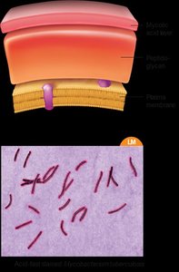
Acid-Fast Staining
Acid-fast staining distinguishes bacteria with waxy, mycolic acid-rich cell walls (e.g., Mycobacterium species). Acid-fast bacteria retain the primary red dye after acid wash, while non–acid-fast cells do not.
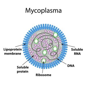
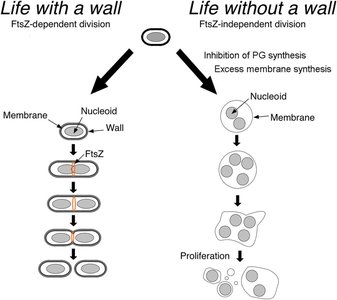
Mycoplasma and L-Forms
Mycoplasma lack a cell wall and instead have a sterol-enriched plasma membrane, making them pleomorphic and resistant to antibiotics targeting cell walls. L-forms are bacteria that can lose their cell wall during their life cycle.
Internal Structures of Prokaryotic Cells
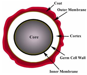
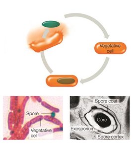
Endospores
Endospores are dormant, highly resistant structures formed by certain bacteria (notably Bacillus and Clostridium genera) in response to harsh conditions. They allow survival through extreme stress and can germinate into vegetative cells when conditions improve.
Nucleoid
The nucleoid is the region within a prokaryotic cell where the single, circular chromosome is located. It is not membrane-bound and is irregularly shaped.
Ribosomes
Prokaryotic ribosomes (70S) are composed of a large (50S) and a small (30S) subunit. They are the site of protein synthesis, essential for cell function and growth.
Cytoskeleton
The prokaryotic cytoskeleton is made of protein filaments that provide structural support, help organize cell division, and direct cell wall construction.
Inclusion Bodies
Inclusion bodies are storage sites for nutrients and other substances. Examples include carboxysomes (carbon fixation) and magnetosomes (magnetic iron accumulation).
Clinical Relevance
Understanding prokaryotic cell structure is essential for clinical microbiology, as it informs the selection of antibiotics and the diagnosis of infections. For example, Gram-negative bacteria are generally more resistant to antibiotics due to their outer membrane, while Gram-positive bacteria are more susceptible to drugs targeting peptidoglycan synthesis.
