 Back
BackIntroduction to Prokaryotic Cells: Structure, Function, and Clinical Relevance
Study Guide - Smart Notes
 Tailored notes based on your materials, expanded with key definitions, examples, and context.
Prokaryotic Cell Basics
Overview of Prokaryotic Domains
Prokaryotic cells are fundamental to life on Earth, having originated approximately 3.8 billion years ago. They are classified into two distinct domains: Bacteria and Archaea.
Bacteria and Archaea are both unicellular and lack a membrane-bound nucleus and organelles.
One key difference: Bacteria have cell walls containing peptidoglycan, while Archaea have pseudopeptidoglycan.
One similarity: Both domains are prokaryotic and share basic cellular structure.
Prokaryotic Cell Structure and Size
Prokaryotic cells are typically small, which allows for efficient nutrient uptake and waste removal.
Most prokaryotes range from 0.2 to 2.0 μm in diameter.
Small size is advantageous for rapid growth and adaptation.
Shapes and Arrangements of Prokaryotes
The shape and arrangement of prokaryotic cells are important for identification and pathogenicity.
Monomorphic bacteria have a single, consistent shape.
Pleomorphic bacteria can vary in shape, enhancing survival and transmission.
Common shapes:
Bacilli (rod-shaped)
Cocci (spherical)
Vibrio (comma-shaped)
Stella (star-shaped)
Coccobacilli (ovoid)
Spirochetes (spiral-shaped)
Arrangements:
Diplococci (paired cocci)
Streptococci (chains of cocci)
Staphylococci (grapelike clusters)
Diplobacilli (paired bacilli)
Streptobacilli (chains of bacilli)
Palisade (clusters of bacilli)
Binary Fission
Prokaryotic cells reproduce primarily by binary fission, a simple and efficient process.
DNA is copied.
Cell grows and chromosomes move to opposite ends.
A septum forms at the midpoint, dividing the cell.
Two daughter cells are produced.
Extracellular Structures
Plasma Membrane Structure and Function
The plasma membrane is a thin, flexible phospholipid bilayer that acts as a selective barrier.
Proteins in the membrane serve as transporters, anchors, receptors, and enzymes.
Site for metabolic reactions, including ATP synthesis.
Selective permeability: allows gases, water, and small noncharged substances to diffuse; larger or charged substances require transport proteins.
Membrane Fluidity
Membrane fluidity is essential for cell function and is influenced by temperature and fatty acid composition.
Warmer temperatures increase fluidity; colder temperatures decrease it.
Unsaturated fatty acids improve fluidity in cold conditions; saturated fatty acids make membranes more rigid.
Archaea Plasma Membranes
Bacterial membranes contain linear fatty acids; archaeal membranes contain long-branched fatty acids.
Some archaea form lipid monolayers for stability in extreme environments.
Cell Wall Structure and Function
Cell walls provide rigidity and protection.
Bacteria: cell wall contains peptidoglycan.
Archaea: cell wall contains pseudopeptidoglycan.
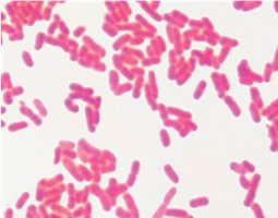
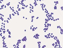
Gram Staining and Acid-Fast Properties
Gram staining is a key clinical tool for classifying bacteria based on cell wall structure.
Gram-positive: thick peptidoglycan layer, stains purple, lacks outer membrane.
Gram-negative: thin peptidoglycan layer, stains red/pink, has outer membrane with lipopolysaccharide.
Acid-fast staining detects mycolic acid in cell walls (e.g., Nocardia, Mycobacterium).
Acid-fast bacteria grow slowly and require long, multidrug therapies.
Clinical Implications of Gram Status
Gram-negative bacteria are harder to kill due to their outer membrane.
Gram-positive bacteria are more sensitive to agents targeting peptidoglycan but retain moisture and resist mechanical stress.
Teichoic acids in Gram-positive cell walls stabilize structure and aid in cell division.
Table: Comparing Gram-Negative and Gram-Positive Bacteria
Feature | Gram-Positive | Gram-Negative |
|---|---|---|
Peptidoglycan Layer | Thick (20–80 nm) | Thin (2–8 nm) |
Outer Membrane | Absent | Present (with lipopolysaccharide) |
Stain Color | Purple | Red/Pink |
Teichoic Acids | Present | Absent |
Porins | Absent | Present |
Resistance to Chemicals | Lower | Higher |
Transport Mechanisms
Passive and Active Transport
Passive transport: no energy required; includes diffusion and osmosis.
Active transport: requires energy; includes primary (ATP-driven), secondary (ion gradient-driven), and phosphotransferase systems.
Diffusion
Simple diffusion: movement of small, noncharged molecules.
Facilitated diffusion: uses membrane proteins (channels/carriers).
Osmosis
Water moves from low solute to high solute concentration.
Isotonic: no net water movement.
Hypertonic: cell loses water, plasmolysis occurs.
Hypotonic: cell gains water, osmotic lysis may occur.
Active Transport Mechanisms
Primary: uses ATP.
Secondary: uses ion gradients (symport and antiport).
Phosphotransferase: transfers phosphate to transported substance (e.g., glucose).
External Structures for Adhesion, Movement, and Protection
Flagella
Flagella are protein filaments for motility, powered by a rotary mechanism.
Gram-positive bacteria have two rings; Gram-negative have four rings for anchoring.
Movement types: chemotaxis (chemical), phototaxis (light), aerotaxis (oxygen).
Arrangements: monotrichous (single), lophotrichous (tuft), amphitrichous (both poles), peritrichous (all over).
Periplasmic flagella (axial filaments) allow spirochetes to move in corkscrew motion.
Fimbriae and Pili
Fimbriae: short, bristle-like, for adhesion and biofilm formation.
Pili: longer, fewer, for adhesion, movement, and gene transfer (conjugation).
Glycocalyx
Sticky carbohydrate layer for adhesion, protection, and resistance to desiccation.
Slime layer: unorganized; capsule: organized and tightly associated.
Intracellular Structures
Nucleoid
Region containing single, circular chromosome.
Ribosomes
70S ribosomes (50S + 30S subunits) synthesize proteins.
Cytoskeleton
Protein filaments provide structure and support.
Inclusion Bodies
Storage granules for substances (e.g., carboxysomes, magnetosomes).
Endospores
Metabolically inactive, highly resistant structures for survival in harsh conditions.
Produced by Bacillus, Clostridium, and Clostridioides genera.
Endospores can survive for extended periods, posing challenges in healthcare.
Sporulation
DNA is copied and packaged with ribosomes and enzymes.
Spore core is surrounded by resistant layers and released.
Visual Summary: Introduction to Prokaryotic Cells
Prokaryotic cells are diverse, efficient, and clinically significant.
Understanding their structure and function is essential for microbiology and healthcare.
Additional info: Some content was inferred and expanded for completeness and clarity, including typical prokaryotic cell sizes, sporulation steps, and clinical implications.
