 Back
Introduction to the Microbial World: Structure, Diversity, and Impact
Study Guide - Smart Notes
 Tailored notes based on your materials, expanded with key definitions, examples, and context.
Chapter 1: The Microbial World
Introduction to Microbiology
Microbiology is the study of microorganisms, which are organisms too small to be seen with the naked eye. This field encompasses the structure, function, diversity, and roles of microbes in various environments, including their impact on health, industry, and the biosphere.
Microbial Communities
Microorganisms rarely exist in isolation; instead, they form complex communities where they interact with each other and their environment. These communities can be found in diverse habitats, from soil and water to the human body.
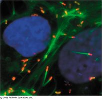
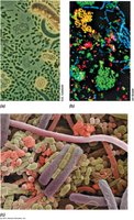
Definition: A microbial community is a group of microorganisms living and interacting in a specific environment.
Importance: Microbial communities are essential for nutrient cycling, ecosystem stability, and the health of plants, animals, and humans.
Example: The human gut microbiome is a complex community that aids in digestion and immune function.
Applications of Microbiology
Microorganisms play critical roles in various sectors, including health, agriculture, industry, and environmental management.
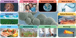
Human and Animal Health: Microbes are involved in disease, immunity, and the production of antibiotics.
Agriculture: Microbes contribute to soil fertility and plant health through nitrogen fixation and decomposition.
Industry and Biotechnology: Microbes are used in fermentation, bioremediation, and the production of biofuels and pharmaceuticals.
Environmental Health: Microbes are essential for wastewater treatment and the cycling of nutrients in ecosystems.
Microbial Cell Structure and Function
Microbial Growth and Colonies
Microbial growth refers to the increase in the number of cells, typically through binary fission. Colonies on solid media originate from a single cell or colony-forming unit (CFU), and ideally, all cells in a colony are genetically identical.
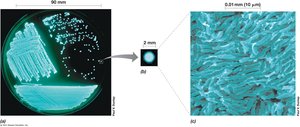
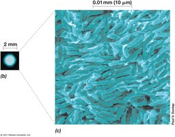
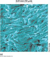
Colony-Forming Unit (CFU): The original cell from which a colony arises.
Genetic Uniformity: Colonies are used to isolate pure cultures for study.
Cell Structure: Prokaryotes vs. Eukaryotes
Microbial cells are classified as prokaryotic (Bacteria and Archaea) or eukaryotic (Eukarya). Their structural differences are fundamental to their biology and classification.
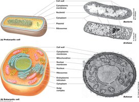
Prokaryotes: Lack a nucleus; DNA is not enclosed by a membrane and is typically circular. Bacteria may have plasmids carrying extra genetic information, such as antibiotic resistance genes.
Eukaryotes: Have a membrane-bound nucleus and organelles such as mitochondria, Golgi apparatus, and endoplasmic reticulum. Metabolism is compartmentalized within organelles.
Cytoplasm: Site of metabolic reactions in all cells.
Properties and Diversity of Microbial Cells
Properties of Microbial Cells
All cells share certain properties, but some features are unique to specific groups of microorganisms.
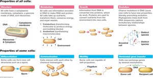
Metabolism: The sum of all chemical reactions in a cell. Includes catabolic (energy-yielding) and anabolic (biosynthetic) pathways.
Growth: Cells grow by assimilating nutrients and dividing to form new cells.
Evolution: Genetic changes allow adaptation and the emergence of new species.
Additional Properties: Some cells can form spores, communicate, move (motility), or exchange genes (horizontal gene transfer).
Cell Size and Volume
Microbial cells vary greatly in size and volume, influencing their physiology and ecological roles.
Organism | Characteristics | Morphology | Size (μm) | Cell Volume (μm³) | Volume Compared to E. coli |
|---|---|---|---|---|---|
Thiomargarita namibiensis | Sulfur chemolithotroph | Coccus in chains | 750 | 200,000,000 | 300,000 |
Epulopiscium fishelsoni | Chemoorganotroph | Rods with tapered ends | 80 × 600 | 150,000,000 | 200,000 |
Escherichia coli | Chemoorganotroph | Rod | 1 × 3 | 1 | 1 |
Mycoplasma pneumoniae | Chemoorganotroph | Pleomorphic | 0.2 × 0.3 | 0.01 | 0.01 |
"Candidatus Pelagibacter ubique" | Chemoorganotroph | Rod | 0.2 × 0.5 | 0.02 | 0.02 |
Additional info: Table simplified for key examples; see original for full list. |

Surface Area-to-Volume Relationships
The ratio of surface area to volume affects nutrient uptake and growth rates in microbial cells. Smaller cells have a higher surface area-to-volume ratio, facilitating faster exchange with the environment.
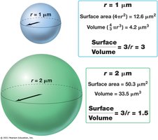
Formula for Surface Area of a Sphere:
Formula for Volume of a Sphere:
Surface Area-to-Volume Ratio:
Cell Morphologies
Microorganisms exhibit a variety of shapes, which can influence their ecological roles and identification.
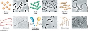
Coccus: Spherical cells
Rod (Bacillus): Cylindrical cells
Spirillum: Spiral-shaped cells
Other Forms: Spirochetes, stalked, filamentous, and pleomorphic cells
Microbial Diversity and Evolution
Microbial Diversity in Size and Shape
Microorganisms vary greatly in their size and shape, which can be observed using different types of microscopy.
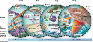
Observation: Light and electron microscopy are used to study microbial morphology and ultrastructure.
Origin and Evolution of Cellular Domains
The history of life on Earth is marked by the evolution of three domains: Bacteria, Archaea, and Eukarya. The Last Universal Common Ancestor (LUCA) is the hypothetical ancestor of all cellular life.
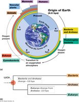
Bacteria and Archaea: Diverged early in Earth's history, both are prokaryotic but differ in molecular and biochemical traits.
Eukarya: Evolved later, characterized by complex cellular organization.
Extremophiles
Some microorganisms, known as extremophiles, thrive in conditions that are extreme for most life forms, such as high temperature, acidity, salinity, or pressure.
Extreme | Descriptive Term | Genus, Species | Domain | Habitat | Minimum | Optimum | Maximum |
|---|---|---|---|---|---|---|---|
High Temperature | Hyperthermophile | Methanopyrus kandleri | Archaea | Hydrothermal vent | 85°C | 98°C | 122°C |
Low pH | Acidophile | Picrophilus oshimae | Archaea | Acidic hot springs | -0.06 | 0.7 | 4 |
High Pressure | Barophile | Photobacterium profundum | Bacteria | Deep sea | 0.1 MPa | 50 MPa | 120 MPa |
High Salt | Halophile | Halobacterium salinarum | Archaea | Salt lakes | 5% | 20% | 32% NaCl |
Additional info: Table simplified for key examples; see original for full list. |

Historical Foundations of Microbiology
Early Microscopy and Discovery of Microbes
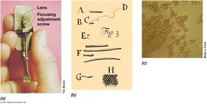
The development of the microscope was crucial for the discovery of microorganisms. Early pioneers like Robert Hooke and Antonie van Leeuwenhoek made significant contributions to the field.
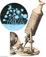
Robert Hooke: First to describe cells in cork and mold using a compound microscope.
Antonie van Leeuwenhoek: First to observe and describe single-celled microorganisms ("animalcules").
Microscopy Techniques
Modern microscopy includes various techniques for observing microbial cells, such as bright-field, fluorescence, and electron microscopy.
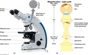
Bright-field Microscopy: Uses visible light to illuminate specimens.
Fluorescence Microscopy: Uses fluorescent dyes to visualize specific structures.
Electron Microscopy: Provides high-resolution images of cell ultrastructure.
Staining and the Gram Stain
Staining techniques, such as the Gram stain, are essential for differentiating bacterial species based on cell wall structure.
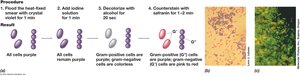
Gram-Positive Bacteria: Retain crystal violet stain and appear purple.
Gram-Negative Bacteria: Do not retain crystal violet and appear red or pink after counterstaining.
Defeat of Spontaneous Generation
Louis Pasteur's experiments disproved the theory of spontaneous generation, demonstrating that life arises from pre-existing life.
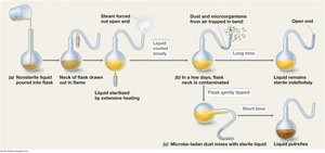
Experiment: Pasteur used swan-necked flasks to show that sterilized broth remained free of microbial growth unless exposed to airborne microorganisms.
Koch's Postulates
Robert Koch established a set of criteria, known as Koch's postulates, to prove the causal relationship between a microorganism and a disease.
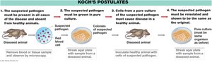
The suspected pathogen must be present in all cases of the disease and absent from healthy animals.
The suspected pathogen must be grown in pure culture.
Cells from a pure culture must cause disease in a healthy animal.
The suspected pathogen must be reisolated and shown to be the same as the original.
Summary
This chapter introduces the microbial world, highlighting the diversity, structure, and essential roles of microorganisms. It also covers foundational concepts in microbial cell biology, the history of microbiology, and the methods used to study microbes. Understanding these basics is crucial for further study in microbiology and its applications in health, industry, and the environment.
