 Back
BackKey Microbial Pathogens and Their Microscopic Identification
Study Guide - Smart Notes
 Tailored notes based on your materials, expanded with key definitions, examples, and context.
Overview of Microbial Pathogens
This guide summarizes important microbial pathogens, their classification, associated diseases, and key microscopic features. It is designed to help students recognize and differentiate between bacteria, viruses, fungi, protozoa, helminths, and arthropod vectors relevant to clinical microbiology.
Bacteria
Mycobacterium tuberculosis (Tuberculosis)
Mycobacterium tuberculosis is the causative agent of tuberculosis (TB). It is an acid-fast bacillus, meaning it retains certain stains even after acid-alcohol decolorization due to its mycolic acid-rich cell wall.
Classification: Acid-fast bacillus (bacteria)
Microscopy: Appears as bright pink rods, often in clumps, when stained with Ziehl-Neelsen stain.
Clinical relevance: Causes pulmonary and extrapulmonary TB, a major global health concern.
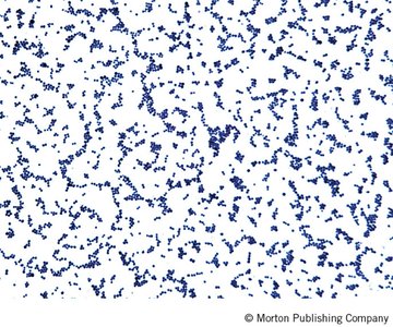
Staphylococcus aureus (Staph Infection)
Staphylococcus aureus is a Gram-positive coccus that forms clusters resembling grapes. It is a common cause of skin infections, abscesses, and systemic diseases.
Classification: Gram-positive staphylococcus (bacteria)
Microscopy: Dark purple or blue clusters after Gram staining.
Clinical relevance: Responsible for a range of infections from mild skin infections to life-threatening sepsis.
Viruses
Epstein-Barr Virus (Mononucleosis)
Epstein-Barr virus (EBV) is a double-stranded DNA virus associated with infectious mononucleosis. It infects B lymphocytes and epithelial cells.
Classification: dsDNA virus
Microscopy: Not typically visualized directly; diagnosis is based on clinical presentation and serology.
Clinical relevance: Causes mononucleosis, characterized by fever, sore throat, and lymphadenopathy.
Helminths
Ascaris lumbricoides (Ascariasis)
Ascaris lumbricoides is a large intestinal roundworm (nematode) causing ascariasis. Eggs are commonly identified in stool samples.
Classification: Nematode (helminth)
Microscopy: Eggs are pale with a darker center and a bumpy surface.
Clinical relevance: Causes gastrointestinal symptoms and may lead to malnutrition.
Taenia solium (Taeniasis)
Taenia solium is a cestode (tapeworm) responsible for taeniasis and cysticercosis in humans.
Classification: Cestode (helminth)
Microscopy: Eggs have a radiating shell pattern (sunburst appearance); adult worms are segmented.
Clinical relevance: Infection occurs via ingestion of undercooked pork containing cysticerci.
Schistosoma mansoni (Schistosomiasis)
Schistosoma mansoni is a trematode (fluke) causing schistosomiasis. Eggs have a characteristic lateral spine.
Classification: Trematode (helminth)
Microscopy: Eggs appear with a prominent spine, often described as resembling a speech bubble.
Clinical relevance: Causes chronic liver and intestinal disease.
Fungi
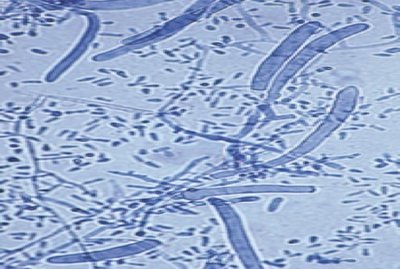
Candida albicans (Candidiasis)
Candida albicans is a yeast that can form pseudohyphae and true hyphae. It is a common cause of mucosal and systemic fungal infections.
Classification: Fungus (yeast)
Microscopy: Appears as oval yeast cells or elongated hyphae.
Clinical relevance: Causes oral thrush, vaginal yeast infections, and invasive candidiasis.
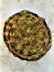
Coccidioides immitis (Valley Fever)
Coccidioides immitis is a dimorphic fungus causing coccidioidomycosis (Valley Fever), primarily in arid regions.
Classification: Fungus
Microscopy: Spherules containing endospores in tissue samples.
Clinical relevance: Causes respiratory illness, which can disseminate in immunocompromised individuals.
Trichophyton rubrum (Dermatophytosis)
Trichophyton rubrum is a dermatophyte fungus responsible for ringworm, athlete's foot, and jock itch (tineas).
Classification: Fungus
Microscopy: Septate hyphae and conidia in skin scrapings.
Clinical relevance: Infects keratinized tissues such as skin, hair, and nails.
Protozoa
Plasmodium spp. (Malaria)
Plasmodium species are protozoan parasites that infect red blood cells, causing malaria. The ring stage is commonly seen in blood smears.
Classification: Protozoan
Microscopy: Ring forms inside red blood cells.
Clinical relevance: Causes cyclical fevers, anemia, and can be life-threatening.
Giardia spp. (Giardiasis)
Giardia species are flagellated protozoa causing gastrointestinal illness. They have a characteristic teardrop shape.
Classification: Protozoan
Microscopy: Teardrop-shaped trophozoites, often pale and difficult to distinguish from the background.
Clinical relevance: Causes diarrhea and malabsorption.
Trichomonas vaginalis (Trichomoniasis)
Trichomonas vaginalis is a flagellated protozoan causing trichomoniasis, a common sexually transmitted infection.
Classification: Protozoan
Microscopy: Motile trophozoites with visible flagella.
Clinical relevance: Causes vaginitis and urethritis.
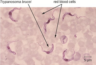
Trypanosoma spp. (Chagas Disease, African Sleeping Sickness)
Trypanosoma species are protozoa that appear as elongated, worm-like organisms in blood smears. They cause diseases such as Chagas disease and African sleeping sickness.
Classification: Protozoan
Microscopy: Slender, undulating forms among red blood cells.
Clinical relevance: Transmitted by insect vectors; can cause severe cardiac and neurological disease.
Arthropods and Vectors
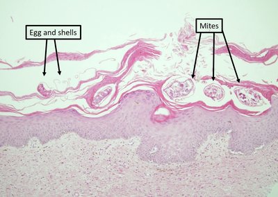
Sarcoptes scabiei var. hominis (Scabies)
Sarcoptes scabiei is a mite that burrows into the skin, causing scabies. Eggs are bean-shaped and found under the skin surface.
Classification: Animal (pathogen, mite)
Microscopy: Mites and eggs visible in skin scrapings.
Clinical relevance: Causes intense itching and rash.
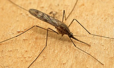
Anopheles spp. (Mosquito Vector)
Anopheles mosquitoes are biological vectors for malaria and other diseases. They are identified by their resting posture and palps as long as the proboscis.
Classification: Animal (biological vector, mosquito)
Microscopy: Not typically examined microscopically for diagnosis; identification is based on morphology.
Clinical relevance: Transmit Plasmodium species and other pathogens.
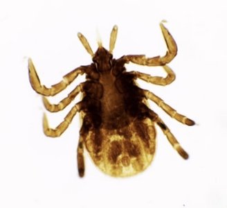
Ixodes spp. (Tick Vector)
Ixodes ticks are vectors for Lyme disease and other tick-borne illnesses. They are recognized by their hard body and mouthparts.
Classification: Animal (biological vector, tick)
Microscopy: Identified by their morphology under low magnification.
Clinical relevance: Transmit Borrelia burgdorferi (Lyme disease) and other pathogens.
Summary Table: Key Microbial Pathogens
Scientific Name | Common Name | Classification | Associated Disease |
|---|---|---|---|
Epstein-Barr virus | None | dsDNA virus | Mononucleosis |
Mycobacterium tuberculosis | None | Acid-fast bacillus (bacteria) | Tuberculosis |
Staphylococcus aureus | None | Gram+ staphylococcus (bacteria) | Staph infection |
Ascaris lumbricoides | Roundworm | Nematode (helminth) | Ascariasis |
Taenia solium | Tapeworm | Cestode (helminth) | Taeniasis |
Schistosoma mansoni | None | Trematode (helminth) | Schistosomiasis |
Candida albicans | Yeast | Fungus | Candidiasis |
Coccidioides immitis | None | Fungus | Valley fever |
Trichophyton rubrum | None | Fungus | Ringworm, athlete's foot, jock itch |
Plasmodium spp. | None | Protozoan | Malaria |
Giardia spp. | None | Protozoan | Giardiasis |
Trichomonas vaginalis | None | Protozoan | Trichomoniasis |
Trypanosoma spp. | None | Protozoan | Chagas disease, African sleeping sickness |
Sarcoptes scabiei var. hominis | Mite | Animal (pathogen) | Scabies |
Anopheles spp. | Mosquito | Animal (biological vector) | Malaria (vector) |
Ixodes spp. | Tick | Animal (biological vector) | Lyme disease (vector) |
