 Back
Back5-5
Study Guide - Smart Notes
 Tailored notes based on your materials, expanded with key definitions, examples, and context.
Koch’s Postulates and the Germ Theory of Disease
Historical Foundations and Importance
The Germ Theory of Disease established that microbes are causative agents of disease. To provide direct evidence linking a specific microbe to a specific disease, Koch’s Postulates were developed in the late 19th century. These postulates remain fundamental to our understanding of infectious diseases, though modern molecular techniques have prompted updates and revisions.
Koch’s Postulate 1: The suspected agent must be present in every case of the disease and absent from healthy organisms.
Koch’s Postulate 2: The suspected pathogen must be isolated and grown in pure culture.
Koch’s Postulate 3: The cultured agent must cause disease when inoculated into a healthy, susceptible host.
Koch’s Postulate 4: The same agent must be reisolated from the diseased experimental host.
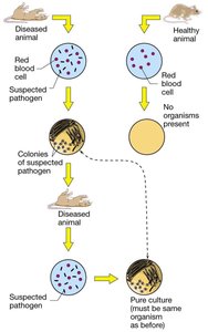
Significance: Koch’s Postulates provide a systematic method for identifying the causative agent of infectious diseases, which is essential for developing prevention and treatment strategies.
Limitations and Modern Updates
Many pathogens do not fulfill all of Koch’s Postulates due to unique biological properties or ethical constraints. For example:
Bacteria that lose or gain plasmids may not be identical upon reisolation (Postulate 4).
Viruses requiring human cells cannot be grown in pure culture (Postulate 2).
Human-only pathogens cannot ethically be tested in healthy human hosts (Postulate 3).
Modern molecular techniques have led to updated criteria, such as those proposed by Fredricks and Relman (1996), which use nucleic acid sequence data to establish causation.
Pathogenicity and Virulence
Definitions and Concepts
Pathogenicity is the ability of a microbe to cause disease, while virulence is a quantitative measure of pathogenicity. Highly virulent microbes cause disease readily, often with a low infectious dose, whereas less virulent microbes require higher numbers to establish infection.
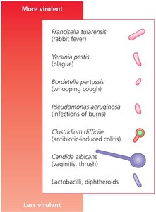
Non-pathogenic organisms do not cause disease.
Virulence factors are molecules produced by pathogens that enhance their ability to cause disease.
Bacterial Virulence Factors
Overview of Virulence Factors
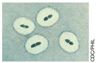
Virulence factors are substances or strategies that enable pathogens to colonize hosts, evade or inhibit the immune response, obtain nutrients, and cause damage. They include enzymes, toxins, and anti-phagocytic factors.
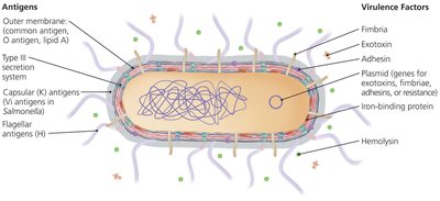
Enzymes (e.g., hyaluronidase, collagenase, coagulase, streptokinase)
Toxins (exotoxins and endotoxins)
Anti-phagocytic factors (e.g., capsules, catalase)
Extracellular Enzymes and Invasion
Extracellular enzymes facilitate infection by breaking down host tissues and promoting invasion.
Hyaluronidase breaks down hyaluronic acid in the extracellular matrix, allowing bacteria to penetrate deeper tissues.
Collagenase degrades collagen in connective tissues, aiding in tissue invasion.
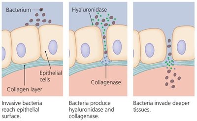
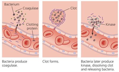
Some enzymes interact with host clotting factors:
Coagulase induces clot formation around bacteria, protecting them from immune cells.
Streptokinase dissolves clots, allowing bacteria to spread.
Examples of Enzyme Virulence Factors
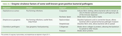
Organism | Disease | Enzyme | Enzyme Activity |
|---|---|---|---|
Staphylococcus aureus | Pus-forming infections | Coagulase | Induces fibrin clotting, protects from immune response |
Streptococcus pyogenes | Scarlet fever, strep throat | Hyaluronidase, Streptokinase | Breaks down ECM, dissolves clots |
Clostridium perfringens | Gas gangrene | Collagenase, Protease | Breaks down collagen and proteins |
Comparison of Staphylococcal Virulence Factors
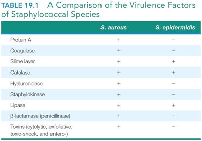
Virulence Factor | S. aureus | S. epidermidis |
|---|---|---|
Protein A | + | – |
Coagulase | + | – |
Slime layer | + | + |
Catalase | + | + |
Hyaluronidase | + | – |
Staphylokinase | + | – |
Lipase | + | + |
β-lactamase | + | – |
Toxins | + | – |
Toxins: Exotoxins and Endotoxins
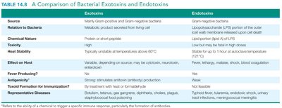
Toxins are molecules that damage host cells or elicit harmful immune responses. They are classified as exotoxins or endotoxins based on their origin and properties.
Property | Exotoxins | Endotoxins |
|---|---|---|
Source | Gram-positive and Gram-negative bacteria | Gram-negative bacteria only |
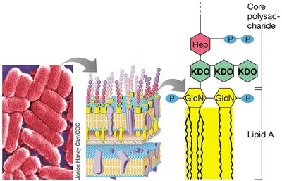
Chemical Nature | Protein or polypeptide | Lipid A of LPS |
Heat Stability | Unstable (destroyed above 60°C) | Stable (withstands autoclaving) |
Toxicity | High | Low (but can be fatal in large amounts) |
Fever Producing | No | Yes |
Antigenicity | Strong | Weak |
Representative Diseases | Diphtheria, tetanus, cholera | Typhoid fever, meningococcemia |
Major Groups of Exotoxins
Cytotoxins: Kill host cells (e.g., hemolysins, leukocidins)
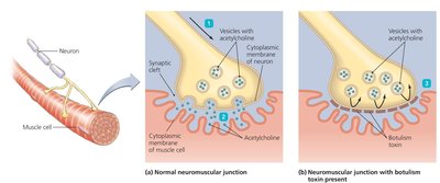
Neurotoxins: Disrupt nerve cell function (e.g., botulinum toxin, tetanus toxin)
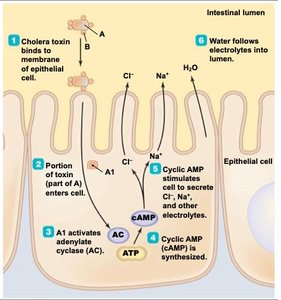
Enterotoxins: Affect the gastrointestinal tract (e.g., cholera toxin)
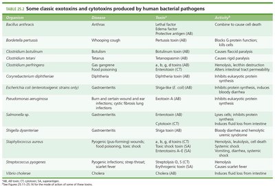
Organism | Toxin | Activity |
|---|---|---|
Bacillus anthracis | Lethal factor, Edema factor | Combine to cause cell death |
Clostridium botulinum | Botulinum toxin | Causes flaccid paralysis |
Vibrio cholerae | Cholera toxin | Induces fluid loss from intestine |
Cytotoxins
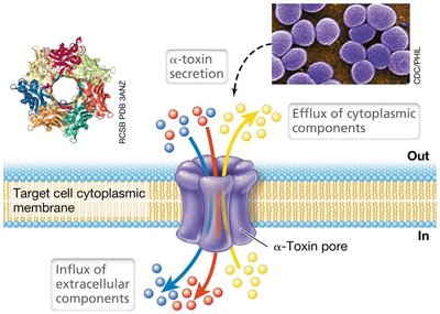
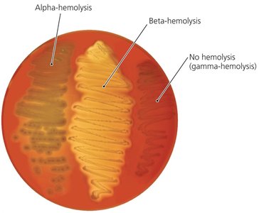
Cytotoxins, such as hemolysins and leukocidins, lyse host cells to release nutrients or evade immune responses. Hemolysins target red blood cells, while leukocidins target white blood cells.
Neurotoxins
Neurotoxins interfere with neurotransmitter release, causing paralysis. Botulinum toxin blocks acetylcholine release, resulting in flaccid paralysis. Tetanus toxin prevents inhibitory neurotransmitter release, causing spastic paralysis.
Enterotoxins
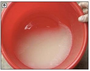
Enterotoxins, such as cholera toxin, disrupt the function of intestinal epithelial cells, leading to massive fluid loss and diarrhea. This facilitates pathogen transmission via contaminated food or water.
Endotoxin (Lipid A of LPS)
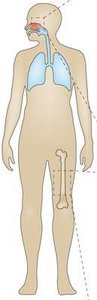
Endotoxin is the lipid A component of lipopolysaccharide (LPS) found in the outer membrane of Gram-negative bacteria. It is released upon bacterial cell lysis and can trigger strong immune responses, including fever, inflammation, and septic shock.
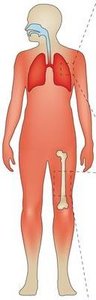
Systemic exposure to endotoxin can cause cytokine storm, shock, and organ failure.
Local infections cause localized inflammation; systemic infections can be fatal.

Anti-phagocytic Factors
Mechanisms of Immune Evasion
Anti-phagocytic factors help bacteria evade destruction by host immune cells. These include:
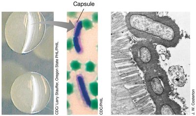
Capsules: Polysaccharide layers that prevent recognition and engulfment by phagocytes.
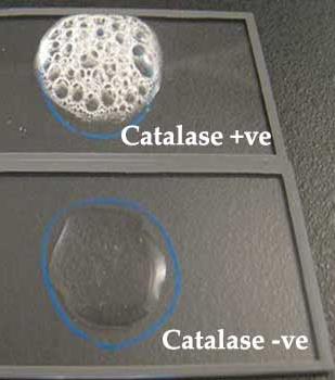
Catalase: Enzyme that neutralizes reactive oxygen species produced by phagocytes.
Toxins and enzymes: Directly attack or kill phagocytes.
Detection of Endotoxin: The LAL Assay
Principle and Application
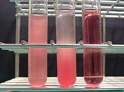
The Limulus amoebocyte lysate (LAL) assay uses blood cells from horseshoe crabs to detect trace amounts of LPS (endotoxin) in pharmaceuticals and medical devices. If LPS is present, the lysate coagulates, indicating contamination.
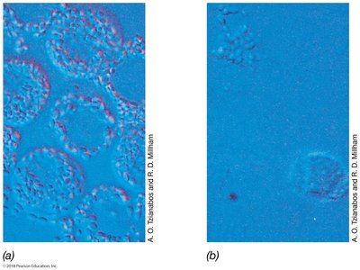
Application: Ensures safety of injectable drugs and medical equipment by confirming absence of endotoxin contamination.
Summary Table: Key Virulence Factors and Their Effects
Virulence Factor | Mechanism | Effect on Host |
|---|---|---|
Hyaluronidase | Degrades hyaluronic acid | Facilitates tissue invasion |
Collagenase | Degrades collagen | Promotes spread through tissues |
Coagulase | Induces clot formation | Protects bacteria from immune cells |
Streptokinase | Dissolves clots | Enables bacterial dissemination |
Exotoxins | Protein toxins secreted by bacteria | Cell death, immune disruption |
Endotoxin (Lipid A) | Component of LPS | Fever, inflammation, shock |
Capsule | Polysaccharide layer | Prevents phagocytosis |
Additional info:
Vehicle-borne transmission (e.g., food, water) is a key mechanism for pathogens producing enterotoxins.
Hemolysins and leukocidins target different cell types: red blood cells and white blood cells, respectively.
