 Back
BackLab Review 7- 12: Differential Stains, Biochemical Tests, and Microbial Control
Study Guide - Smart Notes
 Tailored notes based on your materials, expanded with key definitions, examples, and context.
Lab 6: Differential Stains
Gram Stain
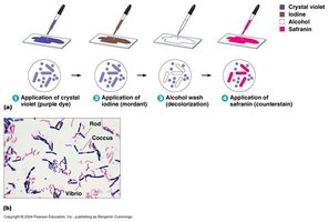
The Gram stain is a fundamental differential staining technique used to classify bacteria into two groups: Gram-positive and Gram-negative, based on differences in their cell wall structure.
Procedure: Involves four main steps: application of crystal violet (primary stain), iodine (mordant), alcohol wash (decolorizer), and safranin (counterstain).
Results: Gram-positive bacteria retain the crystal violet and appear purple; Gram-negative bacteria lose the crystal violet during decolorization and take up safranin, appearing pink/red.
Major Shapes: Bacteria can be rod-shaped (bacilli), spherical (cocci), or spiral (vibrio).
Common Errors:
Over- or under-decolorization with alcohol can lead to incorrect results.
Improper heat fixing, smear thickness, reagent freshness, and culture age can affect outcomes.
Endospore Stain
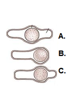
The endospore stain differentiates bacterial endospores from vegetative cells. Endospores are highly resistant structures formed by certain genera, such as Bacillus and Clostridium.
Spore Location: Endospores may be central, terminal, or subterminal within the cell.
Staining: Steam is used to drive malachite green into the endospore; safranin is used as a counterstain for vegetative cells.
Results: Endospores appear green, vegetative cells appear pink.
Acid-Fast Stain
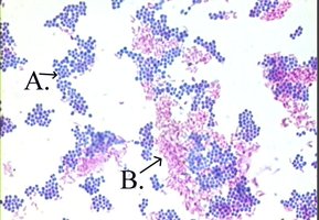
The acid-fast stain is used to identify bacteria with waxy cell walls containing mycolic acid, such as Mycobacterium and Nocardia.
Staining: Steam is used to drive the primary stain into the cell wall.
Results: Acid-fast cells appear pink and often clump together; non-acid-fast cells appear blue.
Lab 8 & 9: Biochemical Tests
Overview of Biochemical Tests
Biochemical tests are used to identify bacteria based on their metabolic properties and enzymatic activities.
Metabolism: The sum of all chemical reactions in an organism.
Indicator: A chemical that changes color to signal a biochemical reaction.
Phenylethyl Alcohol Agar (PEA)
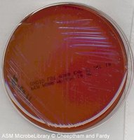
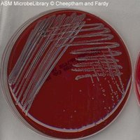
PEA is a selective medium that inhibits Gram-negative bacteria, allowing Gram-positive bacteria to grow. It is often enriched with blood to support fastidious organisms.
Positive Result: Growth indicates Gram-positive bacteria; absence of growth suggests Gram-negative inhibition.
Mannitol Salt Agar (MSA)
MSA is both selective and differential. High salt concentration selects for staphylococci; mannitol fermentation differentiates species.
Indicator: Phenol red turns yellow in acidic conditions due to mannitol fermentation.
Yellow Zone: Indicates mannitol fermentation, typical of Staphylococcus aureus.
Blood Agar Plate (BAP)
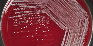
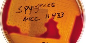
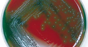
BAP is an enriched and differential medium used to detect hemolytic activity of bacteria.
Types of Hemolysis:
Beta (β) hemolysis: Complete lysis of red blood cells, clear zone around colonies.
Alpha (α) hemolysis: Partial lysis, greenish discoloration.
Gamma (γ) hemolysis: No hemolysis, no change in medium.
Bile Esculin Test
This test identifies bacteria that can hydrolyze esculin in the presence of bile salts. Ferric citrate is the indicator, turning black in the presence of esculetin.
Positive Result: More than half the medium turns black.
Catalase Test
The catalase test detects the presence of the catalase enzyme, which breaks down hydrogen peroxide into water and oxygen (bubbles).
Positive Test: Immediate bubbling upon addition of hydrogen peroxide.
Example: Staphylococcus aureus is catalase positive.
DNAse Test
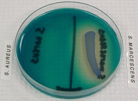
The DNAse test detects the production of the exoenzyme DNAse, which hydrolyzes DNA. Methyl green dye is used as an indicator.
Positive Result: Clear zone around colonies indicates DNA hydrolysis.
Phenol Red Glucose Broths
These broths test for fermentation of glucose and gas production. Phenol red is the pH indicator; a Durham tube captures gas.
Results: Yellow color indicates acid production; gas bubble in Durham tube indicates gas production.
Eosin Methylene Blue (EMB) Agar
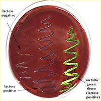
EMB agar is selective for Gram-negative bacteria and differential for lactose fermentation.
Results: Lactose fermenters produce dark colonies with a metallic green sheen; non-fermenters are colorless or pink.
Urease Test
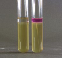
The urease test detects the enzyme urease, which hydrolyzes urea to ammonia and carbon dioxide. Phenol red is the indicator.
Positive Result: Pink color indicates alkaline pH due to ammonia production.
Citrate Test
The citrate test determines if an organism can use citrate as its sole carbon source. Bromthymol blue is the pH indicator.
Positive Result: Blue color indicates alkaline pH from citrate utilization.
SIM Test (Sulfur, Indole, Motility)
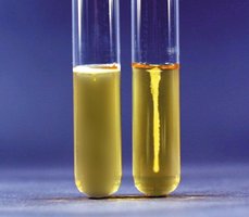
The SIM test is a combination test for sulfur reduction, indole production, and motility.
Sulfur Reduction: Black precipitate indicates H2S production (ferrous ions as indicator).
Indole Production: Red color after addition of Kovac's reagent indicates indole production.
Motility: Cloudy medium indicates motility.
Oxidase Test
The oxidase test detects the presence of cytochrome c oxidase using tetramethyl-p-phenylenediamine as an indicator. A positive result is a color change (usually to dark purple).
Lab 10: Chemical and Antimicrobial Control
Physical and Chemical Control of Microbes
Microbial death is influenced by physical factors (temperature, pH), exposure time, microbial resistance, physiological state, number and location of microbes, presence of organic/inorganic matter, chemical concentration, and mode of action.
Modes of Action: Affect cell wall, cell membrane, proteins, or nucleic acids.
Definitions
Disinfection: Destruction of most microbes on nonliving surfaces.
Bacteriostatic: Inhibits bacterial growth.
Bactericidal: Kills bacteria.
Antisepsis: Destruction of microbes on living tissue.
Antimicrobial Drugs
Ideal Characteristics: Readily available, inexpensive, chemically stable, easily administered, nontoxic, nonallergenic, selectively toxic.
Testing Antimicrobial Efficacy
Disk-Diffusion (Kirby-Bauer): Measures zone of inhibition on Mueller-Hinton agar to determine sensitivity.
Broth Dilution Test: Determines minimum inhibitory concentration (MIC) and minimum bactericidal concentration (MBC).
Lab 11: Analysis of Water
Water Testing
Water is tested for pathogenic microorganisms using coliforms as indicators. Standards vary for drinking, shellfishing, and recreational water.
Coliform: Gram-negative, lactose-fermenting, rod-shaped bacteria (e.g., Escherichia coli).
Multitube Fermentation Test: Includes presumptive, confirmed, and completed tests. If presumptive is negative, no further testing is done.
Lab 11/12: Food Analysis
Food Intoxication vs. Infection
Food Intoxication: Illness caused by ingestion of toxins produced by microbes in food.
Food Infection: Illness caused by ingestion of microbes that then grow in the host.
Example: Salmonella causes food infection, not intoxication.
Hektoen Agar
Used to differentiate E. coli (salmon-orange colonies) from other enteric bacteria.
Additional info: Where images were included, they directly illustrate the described laboratory technique or result. All explanations have been expanded for clarity and academic completeness.
