 Back
Lab Tools and Procedures in Microbiology: The 6 I’s and Microscopy
Study Guide - Smart Notes
 Tailored notes based on your materials, expanded with key definitions, examples, and context.
Lab Tools and Procedures in Microbiology
Challenges of Microbiology
Microbiology involves the study of organisms too small to be seen with the naked eye. These organisms often live in complex associations, requiring special tools and laboratory techniques for isolation and analysis.
Microscopic size: Microbes require magnification for observation.
Complex associations: Microbes interact with each other and their environment, making isolation challenging.
Laboratory study: Microbes must often be separated from their native habitats for controlled study.
The 6 “I”s of Microbiology
Overview of the 6 I’s
The 6 I’s are a systematic approach for studying microorganisms in the laboratory. They include: Inoculation, Incubation, Isolation, Inspection, Information Gathering, and Identification. Proper specimen collection precedes these steps to ensure accurate results.
Specimen Collection: Use aseptic techniques (hand washing, gloves, sterile containers) and proper labeling to avoid contamination.
Common sampling sites: Body fluids, food, water, soil, animals.
Inoculation
Inoculation is the process of introducing a sample into a container of nutrient media to support microbial growth.
Media types: Liquid, solid, or specialized (e.g., eggs).
Tools: Inoculating loops, needles, swabs, syringes.
Purpose: Increases visibility and allows handling of microbes in a controlled environment.
Incubation
Incubation involves placing inoculated media in optimal conditions (temperature, humidity) to promote microbial growth.
Incubator: Maintains controlled environment, typically at 37°C.
Duration: Minimum 18 hours, up to several days.
Purpose: Promotes multiplication and produces visible cultures for further study.
Isolation
Isolation separates individual microbes to obtain pure colonies, each derived from a single cell. This is essential for accurate identification and further testing.
Colony: A mound of cells from a single species.
Purpose: Ensures purity for subsequent analysis.
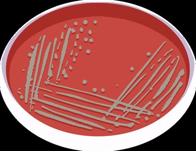
Isolation Techniques
Streak Plate: Dilutes sample across quadrants on a plate to separate colonies.
Pour Plate: Involves diluting sample in melted agar and pouring into plates.
Spread Plate: Dilutes sample in broth, then spreads a drop onto plates.
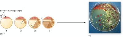
Inspection
Inspection involves observing the physical characteristics of colonies and cells, both macroscopically and microscopically.
Macroscopic: Texture, size, shape, elevation, margin, pigment, and growth requirements (temperature, nutrients).
Microscopic: Cell shape, arrangement, size, and staining reactions (simple and differential stains).
Information Gathering
Information gathering uses various tests to analyze pure cultures for phenotypic, biochemical, genotypic, and immunologic characteristics.
Phenotypic: Observable traits, including enzyme activity and metabolic pathways.
Biochemical: Specialized media and reagents detect enzyme presence (positive/negative results).
Antibiotic Sensitivity: Determines susceptibility to antimicrobial agents, guiding treatment choices.

Genotypic Characteristics
Genotypic analysis examines the genetic material of microbes, often without the need for culture.
DNA Probes: Short DNA sequences that bind to specific microbial DNA, detected via fluorescence microscopy.
PCR (Polymerase Chain Reaction): Amplifies specific DNA segments for rapid detection of pathogens.

Immunologic Characteristics
Immunologic tests detect antigen-antibody reactions in blood or other fluids, aiding in diagnosis and identification.
Antigen: Pathogen component recognized by the immune system.
Antibody: Host molecule that binds to antigens for immune response.
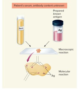
Serological Diagnosis: Patient serum is exposed to known antigens; agglutination indicates presence of specific antibodies.
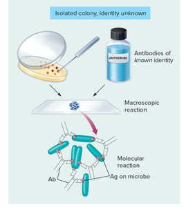
Serotyping: Bacterial colonies are exposed to known antibodies; agglutination identifies the microbe.
Test Properties: Specificity (detects only target antigen/antibody) and sensitivity (detects small amounts).
False Negatives: Microbe present but not detected (e.g., due to variant strains).
False Positives: Microbe not present but test is positive (e.g., due to contamination).
Identification
Identification integrates all collected data to determine the type of microbe. Dichotomous keys are often used to guide the identification process.
Applications: Infection diagnosis, biotechnology, microbial ecology, and research.
The Microscope
Principles of Microscopy
Microscopes are essential for visualizing microorganisms. Two key features are magnification (enlarging objects) and resolution (clarity of detail).
Field of View (FOV): The area visible through the eyepiece; decreases as magnification increases.
Types of Microscopes
Light Microscope: Uses light for illumination, up to 1000X magnification, inexpensive and widely used.
Electron Microscope: Uses electrons for illumination, magnifies up to 1,000,000X, expensive and less accessible.
