 Back
BackLaboratory Methods for Microbiology: Slide Preparation, Smear Technique, and Gram Staining
Study Guide - Smart Notes
 Tailored notes based on your materials, expanded with key definitions, examples, and context.
Laboratory Methods for Microbiology
Preparing a Glass Slide
Proper preparation of glass slides is essential for observing microorganisms under the microscope. Slides must be clean, flat, and correctly labeled to ensure accurate results.
Slide Selection: Use flat slides, not concave, for most microbiological applications.
Labeling: Use a pencil to label each slide with the organism letter (e.g., E, B) for identification.
Slide Storage: Store prepared slides in a slide box to prevent contamination or damage.

Example: Pair Bacillus subtilis (Gram-positive) with Klebsiella pneumoniae (Gram-negative) for comparative staining.
Microbial Cultures and Pairing
Microbiology labs often use paired cultures to compare Gram-positive and Gram-negative bacteria. Each pair consists of one Gram-positive and one Gram-negative organism.
Common Gram-positive: Bacillus subtilis, Staphylococcus epidermidis
Common Gram-negative: Escherichia coli, Klebsiella pneumoniae
Pairing: Each Gram-positive is paired with a Gram-negative for staining and observation.
Smear Preparation from Solid Culture
How to Make a Smear
Creating a bacterial smear is a foundational technique for staining and microscopic examination. The goal is to spread a thin, even layer of bacteria on the slide.
Step 1: Transfer 1-2 drops of water onto the slide and spread thinly.
Step 2: Flame sterilize the inoculating loop before and after each use.
Step 3: Collect a small amount of culture with the loop and mix into the water drop to create a thin smear.
Step 4: Allow the smear to air dry completely before proceeding to staining.
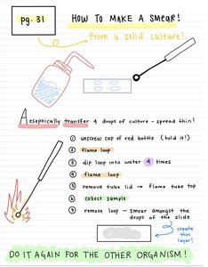
Example: Repeat the smear preparation for each organism being studied.
Gram Staining Procedure
Staining and Heat Fixation
Gram staining differentiates bacteria based on cell wall structure. Proper order and technique are critical for accurate results.
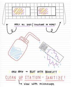
Step 1: Apply all dyes and solutions in the correct order as specified in the protocol.
Step 2: After staining, blot the slide gently with a blotting booklet to remove excess liquid.
Step 3: Heat fix the smear by quickly passing the slide through a flame. This kills the bacteria and adheres them to the slide.
Step 4: Always turn off the Bunsen burner when finished to ensure safety.
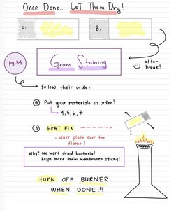
Why Heat Fix? Heat fixing kills bacteria and makes their membranes sticky, ensuring they remain attached during staining.
Microscopy and Slide Observation
Viewing and Clean-Up
After staining, slides are observed under the microscope. Proper clean-up and sanitation are essential to maintain a safe laboratory environment.
Microscopy: Use oil immersion for high-magnification observation of stained bacteria.
Clean-Up: Sanitize the station and properly dispose of used materials after observation.
Culture Media and Inoculation Tools
Types of Culture Media
Different types of media and inoculation tools are used to grow and transfer microorganisms in the lab.
Solid Slant: Used for maintaining stock cultures; inoculated with a loop.
Deep (Stab): Used to test for motility and oxygen requirements; inoculated with a needle.
Broth: Used for growing large numbers of bacteria; inoculated with a loop.
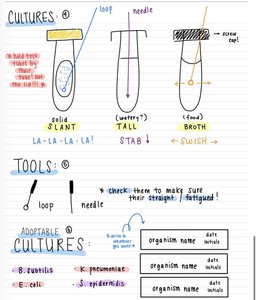
Tools: Always check loops and needles for straightness and fatigue before use.
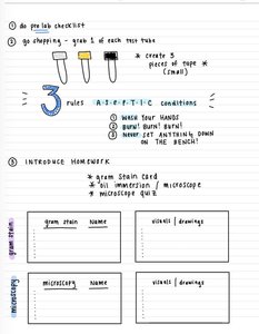
Laboratory Safety and Organization
Rules and Homework
Maintaining aseptic technique and following lab rules is crucial for safety and experimental accuracy.
Wash hands before and after lab work.
Bunsen burner safety: Never leave the flame unattended.
Organization: Label all tubes and slides clearly; never place anything directly on the bench.
Homework: Prepare Gram stain cards, microscope drawings, and complete quizzes as assigned.
Summary Table: Common Laboratory Organisms
Organism | Gram Reaction | Shape | Notes |
|---|---|---|---|
Bacillus subtilis | Positive | Rod | Forms endospores |
Staphylococcus epidermidis | Positive | Coccus | Cluster arrangement |
Escherichia coli | Negative | Rod | Common gut bacterium |
Klebsiella pneumoniae | Negative | Rod | Capsule-forming |
Additional info: This table summarizes the main organisms used in basic microbiology labs for Gram staining and culture techniques.
