 Back
BackLaboratory Techniques in Microbiology: The Five I’s, Media, and Microscopy
Study Guide - Smart Notes
 Tailored notes based on your materials, expanded with key definitions, examples, and context.
Tools of the Laboratory: The Five I’s of Microbiology
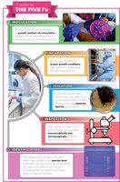
The Five I’s: Overview
The Five I’s are fundamental steps in microbiological laboratory practice, guiding the process from sample collection to microbial identification. Each step is essential for the successful cultivation, observation, and analysis of microorganisms.
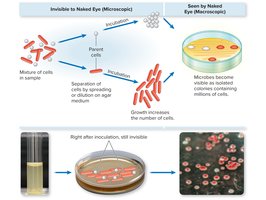
Inoculation: Introduction of a sample (inoculum) into a nutrient medium to initiate microbial growth.
Incubation: Placement of inoculated media in controlled environmental conditions to promote microbial multiplication.
Isolation: Separation of individual microbial species from a mixed sample to obtain pure cultures.
Inspection: Examination of cultures for macroscopic and microscopic characteristics.
Identification: Determination of the microbial species using biochemical, immunological, and genetic methods.
Microbial Cultures: Types and Conditions
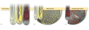
Pure, Mixed, and Contaminated Cultures
Microbial cultures can be classified based on their composition and purity. Understanding these distinctions is crucial for accurate laboratory analysis.
Pure Culture: Contains only one species of microorganism.
Mixed Culture: Contains two or more identified species.
Contaminated Culture: Contains unwanted organisms due to accidental introduction.
Microbiological Media: Physical States and Composition
Physical States of Media
Microbiological media are formulated in different physical states to suit various experimental needs. The choice of media affects microbial growth and observation.
Liquid Media: Used for propagation and biochemical testing.
Semisolid Media: Useful for motility studies and certain biochemical reactions.
Solid/Reversible to Liquid Media: Essential for isolation and colony formation; agar is a common solidifying agent.
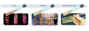
Agar is a complex polysaccharide derived from Gelidium and is not digestible by most microbes. It solidifies at 42°C and liquefies at 100°C.
Media Types: Enriched, Selective, and Differential
Enriched Media
Enriched media contain additional nutrients to support the growth of fastidious organisms, often used in clinical settings to encourage pathogen growth.
Blood Agar: Contains sheep’s blood; used to detect hemolysis.
Chocolate Agar: Contains cooked blood; supports growth of Neisseria species.
Selective and Differential Media
Selective media inhibit the growth of certain microbes while favoring others. Differential media allow multiple species to grow but display visible differences based on biochemical reactions.
Selective Media: Example: MacConkey agar suppresses gram-positive bacteria.
Differential Media: Example: MacConkey agar differentiates lactose fermenters by color change.
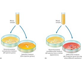
A medium can be both selective and differential, providing both inhibition and differentiation in a single platform.
Miscellaneous Media and Their Purposes
Reducing, Transport, and Fermentation Media
Specialized media serve unique purposes in microbiological analysis.
Reducing Media: Absorbs oxygen, supporting anaerobic bacteria.
Transport Media: Preserves specimens for delayed analysis.
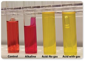
Carbohydrate Fermentation Media: Contains sugars and pH indicators to detect fermentation and gas production.
Isolation Techniques in Microbiology
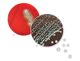
Principles and Methods of Isolation
Isolation is based on separating individual cells to form colonies, which are macroscopic clusters derived from a single cell. This is essential for obtaining pure cultures.
Requirements: Firm surface medium, Petri dish, inoculating loop.
Colony: A visible mound of cells arising from one cell.
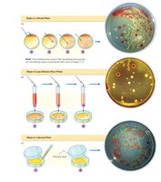
Methods for Isolating Bacteria
Several techniques are used to isolate bacteria, each with specific advantages.
Streak Plate: Sequential spreading to obtain isolated colonies.
Loop Dilution/Pour Plate: Serial dilution and plating for colony separation.
Spread Plate: Even distribution of inoculum across the surface.
Microscopy: Principles and Applications
Size of Macroscopic vs. Microscopic Organisms
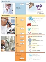
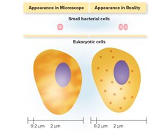
Microorganisms vary greatly in size, requiring different tools for observation. The metric system is used for measurement, ranging from centimeters to nanometers.
Macroscopic: Centimeters and meters (e.g., louse, bread mold).
Microscopic: Millimeters, micrometers, nanometers (e.g., bacteria, viruses, DNA).
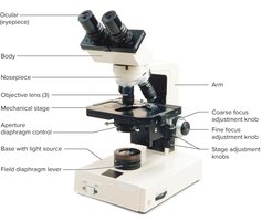
Student Laboratory Microscope: Structure and Function
The compound light microscope is a fundamental tool in microbiology, consisting of several key components for magnification and resolution.
Ocular (eyepiece): Magnifies the image.
Objective lenses: Provide primary magnification.
Stage: Holds the specimen.
Focus knobs: Adjust clarity.
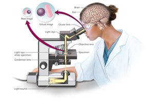
Pathway of Light and Image Formation
Light passes through the specimen, objective lens, and ocular lens, forming a real image and then a virtual image for observation.
Real Image: Formed by the objective lens.
Virtual Image: Formed by the ocular lens.
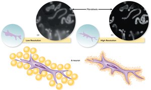
Resolution and Its Importance
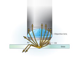
Resolution, or resolving power, is the ability to distinguish two adjacent objects. It is influenced by the wavelength of light and the use of immersion oil.
Human Eye: 0.2 mm
Light Microscope (oil immersion): 0.2 μm
Formula:
Contrast in Microscopy
Contrast is determined by the refractive index difference between specimen and medium. It can be enhanced by adjusting light or using stains and special lenses.
Refractive Index: Degree of light bending.
Iris Diaphragm: Controls light intensity.
Phase-Contrast Microscope: Enhances contrast without staining.
Types of Microscopy
Bright-Field Microscopy
Bright-field microscopy is the most common technique, suitable for both live and stained specimens. It relies on light transmission through the sample.

Dark-Field Microscopy
Dark-field microscopy uses a special condenser to illuminate specimens against a dark background, ideal for observing live, unstained cells.
Phase-Contrast Microscopy
Phase-contrast microscopy exploits differences in cell density to enhance internal detail, useful for observing intracellular structures and motility.
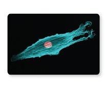
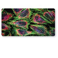
Fluorescence Microscopy
Fluorescence microscopy uses UV light and fluorescent dyes to visualize specific structures or organisms, valuable in diagnostics and research.
Confocal Microscopy
Confocal microscopy employs laser scanning to produce sharp, focused images at various depths, often used with fluorescent stains.
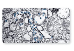
Transmission Electron Microscopy (TEM)
TEM provides detailed views of internal cell structures by transmitting electrons through thinly sectioned specimens.
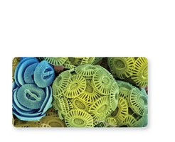
Scanning Electron Microscopy (SEM)
SEM creates three-dimensional images of specimen surfaces by scanning with electrons, offering dramatic and realistic visualization.
Specimen Preparation and Staining Techniques
Fresh, Living Preparations
Wet mounts and hanging drop mounts allow observation of living cells in their natural state, maintaining viability and motility.
Wet Mount: Drop of culture on slide with cover slip.
Hanging Drop: Drop suspended from cover slip over depression slide, sealed with Vaseline.

Staining Methods: Simple, Differential, and Special
Staining enhances visibility and differentiation of microbial cells. Dyes can be basic (positive charge) or acidic (negative charge).
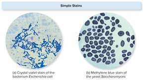
Simple Stains: Use a single dye to reveal cell shape, size, and arrangement.
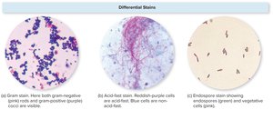
Differential Stains: Use multiple dyes to distinguish cell types or structures (e.g., Gram, acid-fast, endospore stains).
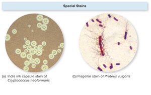
Special Stains: Highlight specific cell parts (e.g., capsule, flagella).
Summary Table: Media Types and Their Purposes
Media Type | Purpose | Example |
|---|---|---|
General-purpose | Grow broad spectrum of microbes | Nutrient agar |
Enriched | Support fastidious organisms | Blood agar, chocolate agar |
Selective | Suppress unwanted microbes | MacConkey agar |
Differential | Distinguish between species | MacConkey agar, blood agar |
Reducing | Grow anaerobes | Thioglycollate broth |
Transport | Preserve specimens | Stuart's transport medium |
Assay | Test antimicrobial effectiveness | Mueller-Hinton agar |
Enumeration | Count organisms | Plate count agar |
