 Back
BackMeasuring and Analyzing Microbial Systems: Culture-Dependent and Culture-Independent Methods
Study Guide - Smart Notes
 Tailored notes based on your materials, expanded with key definitions, examples, and context.
Taking the Measure of Microbial Systems
Introduction
Understanding microbial communities requires a combination of culture-dependent and culture-independent methods. These approaches allow microbiologists to isolate, identify, and analyze the diversity, abundance, and function of microorganisms in various environments.
Culture-Dependent Analyses of Microbial Communities
Enrichment Culture Microbiology
Enrichment culture techniques are used to isolate specific microorganisms from complex samples by providing selective conditions that favor the growth of target organisms while inhibiting others.
Inoculum: The initial sample containing a mixture of microorganisms (e.g., soil, feces, water).
Isolation: The process of separating individual populations from a mixed community.
Enrichment Cultures: Manipulate medium and incubation conditions (nutrients, temperature, pH, oxygen) to select for desired organisms.
Limitations: Enrichment cultures can demonstrate the presence of an organism but cannot prove its absence or ecological importance.
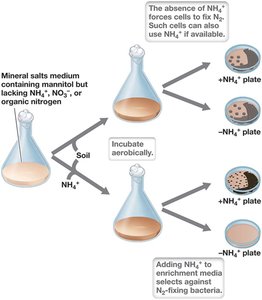
Enrichment Culture Outcomes and Bias
Successful enrichment requires appropriate resources and conditions for target organisms.
Lab cultures often favor fast-growing, minor community members due to higher nutrient concentrations than in nature (enrichment bias).
Dilution of inoculum can help eliminate rapidly growing 'weed' species.
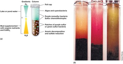
Winogradsky Column: Artificial Microbial Ecosystem
The Winogradsky column is a classic tool for studying microbial ecology, providing gradients of oxygen, light, and nutrients to enrich diverse microbial populations.
Simulates natural environments with layered microhabitats for algae, phototrophs, sulfur-reducers, and anaerobes.
Used to study soil and aquatic microorganisms.
Classical Procedures for Isolating Microbes
Obtaining pure cultures is essential for physiological and molecular studies.
Pure Culture: Contains only one kind of microorganism.
Streak Plate: Technique to isolate single colonies, which are then re-streaked for purity.

Most-Probable-Number (MPN) Technique
The MPN method estimates the number of viable microorganisms in a sample by serial dilution and observation of growth.
Serial 10-fold dilutions in liquid medium.
Used for food, wastewater, and environmental samples.
Growth in a certain dilution indicates at least that many cells per mL in the original sample.

Selective Single-Cell Isolation: Laser Tweezers, Flow Cytometry, Microfluidics
Laser Tweezers: Use focused laser beams to physically separate individual cells for culture, especially slow-growing species.
Flow Cytometry: Counts and sorts cells based on size, shape, or fluorescence as they pass through a detector.

High-Throughput Culture and Microfluidic Devices
Modern techniques allow the isolation and cultivation of single cells in microtiter plates or microfluidic devices, enabling the study of slow-growing or rare microbes.
Microtiter plates allow manipulation of culture conditions for each well.
Microfluidic devices can confine thousands of single cells in nanoliter wells, promoting microcolony growth.
Example: Isolation of Pelagibacter, the most abundant marine bacterium.


Culture-Independent Microscopic Analyses of Microbial Communities
Fluorescent Staining for Enumeration
Fluorescent dyes such as DAPI, acridine orange (AO), and SYBR Green I are used to stain nucleic acids for counting microorganisms under UV light.
DAPI: Stains DNA, fluoresces bright blue.
AO: Stains DNA/RNA, fluoresces orange or greenish orange.
SYBR: Stains DNA, fluoresces green.
Cannot distinguish live from dead cells.

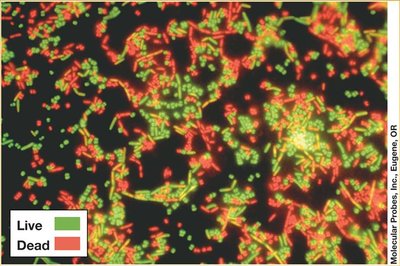
Viability Stains
Viability stains differentiate live and dead cells based on membrane integrity using two dyes: one for live cells (green) and one for dead cells (red).
Useful for assessing cell viability in environmental samples.
Background staining may occur in complex samples.
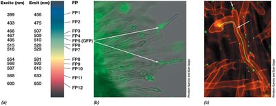
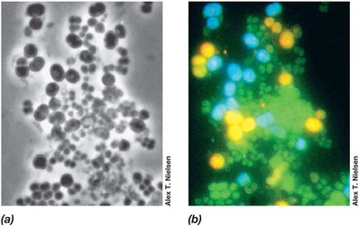
Green Fluorescent Protein (GFP) and Reporter Genes
GFP can be genetically engineered into cells to make them autofluorescent, allowing tracking of live bacteria and gene expression in situ.
Acts as a reporter gene to indicate promoter activity.
Used to study microbial processes such as infection or root colonization.
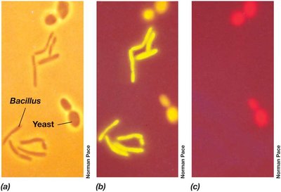
Fluorescence In Situ Hybridization (FISH)
FISH uses fluorescently labeled nucleic acid probes to identify and quantify specific microorganisms in their natural context.
Probes hybridize to rRNA sequences, allowing phylogenetic identification.
Can distinguish between closely related taxa and visualize spatial organization in communities.
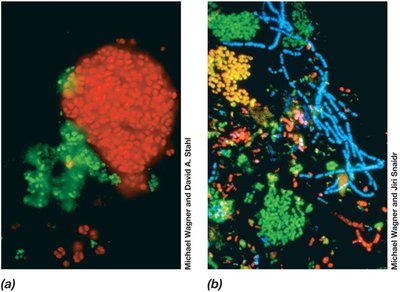
FISH in Environmental and Clinical Applications
FISH can employ multiple probes to analyze complex communities, such as activated sludge in wastewater treatment, and is used in microbial ecology, food industry, and diagnostics.
Environmental Multi-Omics: Genomics, Transcriptomics, Proteomics, Metabolomics
Integration of Omics Approaches
Multi-omics integrates genomic, transcriptomic, proteomic, and metabolomic data to provide a comprehensive understanding of microbial community function and response to environmental change.
Genomics: Analysis of the total gene pool (metagenomics) reveals phylogenetic and metabolic diversity.
Transcriptomics: Metatranscriptomics analyzes community RNA to reveal gene expression patterns.
Proteomics: Metaproteomics measures the diversity and abundance of proteins.
Metabolomics: Comprehensive analysis of metabolites produced by the community.
Environmental Genomics (Metagenomics)
Metagenomics involves cloning and sequencing DNA from environmental samples to detect as many genes as possible, including those not amplified by standard PCR primers.
Provides a snapshot of the gene pool and allows discovery of new genes.
Links genes to specific microbial taxa (phylotypes).
Stable Isotopes and Stable Isotope Probing (SIP)
Stable Isotopes in Microbial Ecology
Stable isotopes (e.g., 12C, 13C, 32S, 34S) are used to study microbial transformations in nature through isotope fractionation.
Enzymes often preferentially incorporate lighter isotopes (e.g., 12C).
Isotopic composition of organic matter reveals past biological activity.
Stable Isotope Probing (SIP)
SIP links specific metabolic activity to microbial diversity by feeding a community a substrate labeled with a stable isotope (e.g., 13C). Organisms that metabolize the substrate incorporate the isotope into their DNA, which can then be separated and analyzed.
DNA with 13C is separated from 12C-DNA by density gradient centrifugation.
Enables identification of active organisms in situ.
Summary Table: Culture-Dependent vs. Culture-Independent Methods
Method | Purpose | Key Features |
|---|---|---|
Enrichment Culture | Isolate specific microbes | Selective media, incubation conditions |
Streak Plate | Obtain pure cultures | Isolation of single colonies |
MPN Technique | Estimate cell numbers | Serial dilution, growth observation |
Laser Tweezers/Flow Cytometry | Single-cell isolation/sorting | Physical separation, fluorescence sorting |
Microfluidics/High-Throughput | Isolate rare/slow-growing species | Single-cell cultivation in microwells |
Fluorescent Staining | Enumerate cells | DAPI, AO, SYBR stains; nonspecific |
Viability Stains | Live/dead differentiation | Dual dyes, membrane integrity |
FISH | Identify/quantify taxa | rRNA probes, phylogenetic specificity |
Multi-Omics | Functional analysis | Genomics, transcriptomics, proteomics, metabolomics |
Stable Isotope Probing | Link function to identity | Isotope-labeled substrates, DNA analysis |
