 Back
BackMetabolic Tests in Microbiology: Study Guide
Study Guide - Smart Notes
 Tailored notes based on your materials, expanded with key definitions, examples, and context.
Metabolic Tests in Microbiology
Mannitol Salt Agar Test
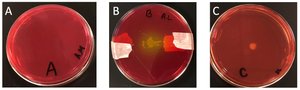
The Mannitol Salt Agar (MSA) Test is used to differentiate and select bacteria based on their ability to tolerate high salt concentrations and ferment mannitol. It is particularly useful for identifying Staphylococcus species.
Selective property: Only bacteria that can grow in high salt (7.5% NaCl) will survive.
Differential property: Bacteria that ferment mannitol produce acid, turning the media yellow due to the phenol red indicator.
Example: Staphylococcus aureus ferments mannitol, causing a yellow color change, while Staphylococcus epidermidis does not.
Oxidase Test
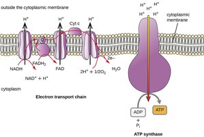
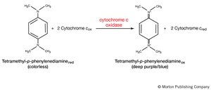
The Oxidase Test detects the presence of cytochrome c oxidase, an enzyme involved in the electron transport chain of aerobic respiration. This test is important for distinguishing between different groups of bacteria, such as Pseudomonas (oxidase positive) and Enterobacteriaceae (oxidase negative).
Principle: If cytochrome c oxidase is present, the electron donor reagent (tetramethyl-p-phenylenediamine) is oxidized and turns purple.
Interpretation: A color change to purple indicates a positive result; no color change indicates a negative result.
Example: Pseudomonas aeruginosa is oxidase positive.
Catalase Test
The Catalase Test identifies bacteria that produce the enzyme catalase, which breaks down hydrogen peroxide (H2O2) into water and oxygen. This is a key test for distinguishing between aerobic and anaerobic bacteria.
Principle: Catalase converts hydrogen peroxide into water and oxygen, producing bubbles.
Interpretation: Bubbling upon addition of hydrogen peroxide indicates a positive result.
Example: Staphylococcus species are catalase positive, while Streptococcus species are catalase negative.

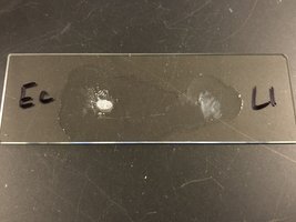
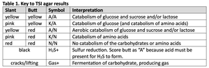
TSI (Triple Sugar Iron) Agar Test
The TSI Agar Test is used to differentiate bacteria based on their ability to ferment glucose, lactose, and sucrose, and to reduce sulfur. The test uses phenol red as a pH indicator.
Fermentation: Acid production turns the media yellow; alkaline conditions turn it pink.
Sulfur reduction: Black precipitate indicates H2S production.
Gas production: Cracks or lifting in the agar indicate gas formation.
Slant | Butt | Symbol | Interpretation |
|---|---|---|---|
yellow | yellow | A/A | Catabolism of glucose and sucrose and/or lactose |
pink | yellow | K/A | Catabolism of glucose (and catabolism of amino acids) |
yellow | red | A/N | Aerobic catabolism of glucose and sucrose and/or lactose |
pink | red | K/N | Catabolism of amino acids |
red | red | N/N | No catabolism of the carbohydrates or amino acids |
black | H2S+ | Sulfur reduction. Score butt as "A" because acid must be present for H2S to form. | |
cracks/lifting | Gas+ | Fermentation of carbohydrate, producing gas |
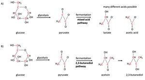
MR-VP Test
The Methyl Red (MR) and Voges-Proskauer (VP) Tests detect different fermentation pathways in bacteria. The MR test identifies mixed acid fermentation, while the VP test detects the production of acetoin via the 2,3-butanediol pathway.
MR Test: Positive result indicates mixed acid fermentation (stable acids).
VP Test: Positive result indicates production of acetoin and 2,3-butanediol.
Example: Escherichia coli is MR positive, VP negative; Enterobacter is MR negative, VP positive.
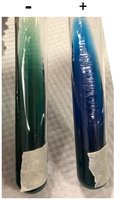
Citrate Test
The Citrate Test determines whether bacteria can use citrate as a sole carbon source. The test uses Simmons citrate agar, which contains bromothymol blue as a pH indicator.
Principle: Utilization of citrate increases pH, turning the media from green to blue.
Interpretation: Blue color indicates a positive result; green indicates negative.
Example: Enterobacter species are citrate positive.
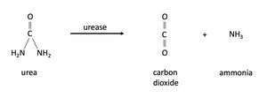
Urease Test
The Urease Test identifies bacteria that produce the enzyme urease, which hydrolyzes urea into ammonia and carbon dioxide. This test is important for identifying Proteus species.
Principle: Ammonia production increases pH, turning the media pink.
Interpretation: Pink color indicates a positive result; no color change indicates negative.
Example: Proteus vulgaris is urease positive.
Nitrate Test
The Nitrate Test determines the ability of bacteria to reduce nitrate to nitrite or further to nitrogen gas (denitrification). This test is useful for differentiating members of the Enterobacteriaceae family.
Principle: Reduction of nitrate is detected by adding reagents after incubation.
Interpretation: Red color after reagents indicates nitrate reduction; gas formation indicates denitrification.
Example: Escherichia coli reduces nitrate to nitrite.
SIM Test: Sulfur, Indole, Motility
The SIM Test is a combination test that assesses three bacterial properties: sulfur reduction, indole production, and motility.
Sulfur reduction: Black precipitate indicates H2S production.
Indole production: Addition of Kovac's reagent produces a red ring if indole is present.
Motility: Growth away from the stab line indicates motility.
Example: Proteus vulgaris is positive for all three tests.
Additional info: These metabolic tests are essential for identifying and differentiating bacterial species in clinical and environmental microbiology. They are commonly used in laboratory settings to assess microbial physiology and metabolic capabilities.
