 Back
Microbial Cell Structure and Function: Bacterial Cell Envelopes and Membranes
Study Guide - Smart Notes
 Tailored notes based on your materials, expanded with key definitions, examples, and context.
Microbial Cell Structure and Function
Overview of Bacterial Cell Structure
Bacterial cells are fundamental units of life in microbiology, characterized by their unique cellular architecture. Understanding their structure is essential for grasping microbial physiology, taxonomy, and pathogenesis.
Cell Envelope: Composed of the cytoplasmic membrane, cell wall, and, in some cases, an outer membrane.
Cytoplasm: Contains ribosomes, DNA, and various inclusions.
Species Naming: The binomial nomenclature (e.g., Escherichia coli) is used for scientific identification.
Biosafety Levels: Laboratory strains (e.g., E. coli K12) are handled at BSL1, while highly pathogenic microbes (e.g., Ebola) require BSL4 containment.
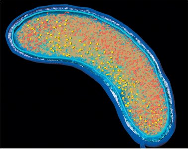
Cytoplasmic Membrane Structure
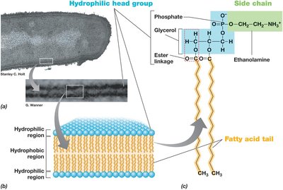
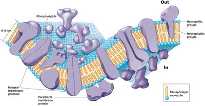
The cytoplasmic membrane is a selectively permeable barrier essential for cellular integrity and metabolic function. It is composed of a phospholipid bilayer with embedded proteins.
Phospholipid Bilayer: Consists of hydrophilic head groups and hydrophobic fatty acid tails.
Integral and Peripheral Proteins: Facilitate transport, signaling, and enzymatic activity.
Membrane Functions: Include permeability barrier, protein anchoring, and energy conservation.
Archaeal Membrane Architecture
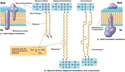
Archaea possess unique membrane lipids distinct from bacteria and eukaryotes, contributing to their ability to thrive in extreme environments.
Ether Linkages: Archaeal membranes contain ether-linked lipids, unlike the ester linkages in bacterial membranes.
Isoprenoid Chains: Provide stability and flexibility.
Monolayer and Bilayer Structures: Some archaea have monolayer membranes for enhanced stability.
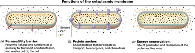
Functions of the Cytoplasmic Membrane
The cytoplasmic membrane is central to cellular homeostasis, energy production, and transport.
Permeability Barrier: Prevents leakage and controls entry/exit of solutes.
Protein Anchor: Site for proteins involved in transport and chemotaxis.
Energy Conservation: Generates and dissipates the proton motive force (pmf), crucial for ATP synthesis.
Transport Across Membranes
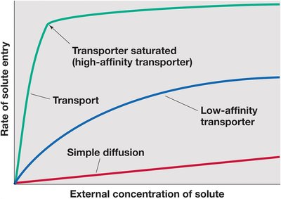
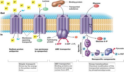
Transport systems are vital for nutrient uptake and waste removal. They include simple diffusion, facilitated transport, and active transport mechanisms.
Simple Diffusion: Movement of solutes down their concentration gradient.
Transporters: Proteins that facilitate solute entry, including high- and low-affinity types.
ABC Transporters: Use ATP to drive transport against gradients.
Group Translocation: Chemical modification of transported molecules (e.g., phosphorylation).
Bacterial Cell Wall Types
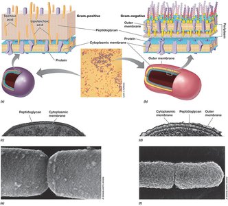
Bacterial cell walls provide structural support and protection. The Gram stain differentiates bacteria based on cell wall composition.
Gram-Positive: Thick peptidoglycan layer, retains crystal violet stain (purple).
Gram-Negative: Thin peptidoglycan layer, outer membrane, loses crystal violet, retains safranin (pink).
Acid-Fast: Rich in mycolic acids, resistant to Gram stain.
Cell-Wall-Deficient: Mycoplasma and L-forms lack peptidoglycan.
S-Layers: Protein or glycoprotein arrays, common in Archaea.
Chlamydial Anomaly: Unique protein network, lacks detectable peptidoglycan.
Peptidoglycan Structure and Function
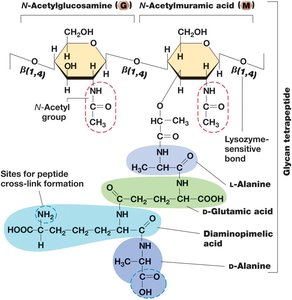
Peptidoglycan is a polymer of sugars and amino acids forming a mesh-like layer outside the plasma membrane. It is essential for cell shape and resistance to osmotic pressure.
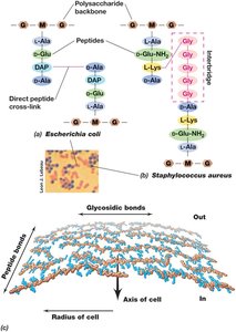
Glycan Tetrapeptide: Repeating units of N-acetylglucosamine (G) and N-acetylmuramic acid (M) cross-linked by peptides.
Lysozyme Sensitivity: Lysozyme cleaves glycosidic bonds, leading to cell lysis.
Peptide Cross-Linking: Provides structural integrity.
Gram-Negative Cell Envelope
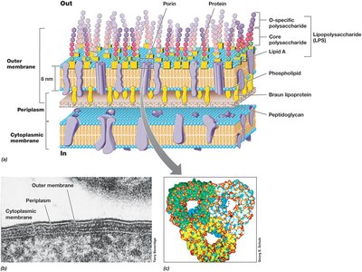
Gram-negative bacteria possess a complex cell envelope with an outer membrane, periplasmic space, and thin peptidoglycan layer.
Outer Membrane: Contains lipopolysaccharide (LPS), porins, and proteins.
Periplasm: Space between the outer and cytoplasmic membranes.
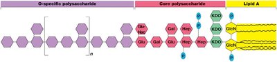
LPS: Composed of O-specific polysaccharide, core polysaccharide, and lipid A.
Gram-Positive Cell Wall
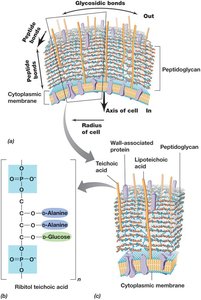
Gram-positive bacteria have a thick peptidoglycan layer with teichoic acids, which contribute to cell wall rigidity and function.
Teichoic Acids: Polymers of glycerol or ribitol, linked to peptidoglycan.
Lipoteichoic Acids: Anchored in the cytoplasmic membrane.
Antibiotic Action on Cell Walls
Antibiotics such as penicillin target peptidoglycan synthesis, making them effective against actively growing Gram-positive bacteria. Gram-negative bacteria are protected by their outer membrane.
Lysozyme: Destroys peptidoglycan by cleaving glycosidic bonds.
Penicillin: Inhibits peptide cross-linking during cell wall synthesis.
Broad Spectrum Antibiotics: Target protein synthesis at the ribosome.
Summary Table: Gram-Positive vs. Gram-Negative Cell Envelope
Feature | Gram-Positive | Gram-Negative |
|---|---|---|
Peptidoglycan Thickness | Thick | Thin |
Outer Membrane | Absent | Present |
Teichoic Acids | Present | Absent |
Lipopolysaccharide (LPS) | Absent | Present |
Stain Color (Gram Stain) | Purple | Pink |
Additional info: The notes above expand on the original content by providing definitions, examples, and academic context for each structural feature and function. Images included are directly relevant to the explanation of each paragraph, visually reinforcing the concepts described.
