 Back
BackMicrobial Cell Structure and Function: Cell Envelope and Membrane Transport
Study Guide - Smart Notes
 Tailored notes based on your materials, expanded with key definitions, examples, and context.
Cell Envelope & Biological Membranes
Definition and Components
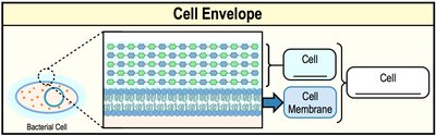
The cell envelope encompasses all layers that surround the cell, including the cell membrane and, in many cases, the cell wall. The composition of the cell envelope varies among different cell types, but all cells possess a cell membrane as a fundamental component.
Cell Envelope: All structural layers surrounding the cell (e.g., membranes, cell walls).
Cell Membrane: Always included in the cell envelope; present in all cells.
Cell Wall: Present in many, but not all, cells (e.g., most bacteria, some eukaryotes).
Biological Membranes and the Fluid Mosaic Model
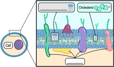
Biological membranes are primarily composed of phospholipids, which are amphipathic molecules, and other embedded molecules such as proteins and cholesterol. The fluid mosaic model describes membranes as dynamic structures with proteins and lipids moving laterally within the bilayer.
Phospholipid Bilayer: The fundamental structure of biological membranes.
Fluid Mosaic Model: Membranes are fluid and contain a mosaic of proteins and lipids.
Proteins: Constitute 20–80% of membrane mass and are mobile within the membrane.
Bacterial, Eukaryotic, and Archaeal Membranes
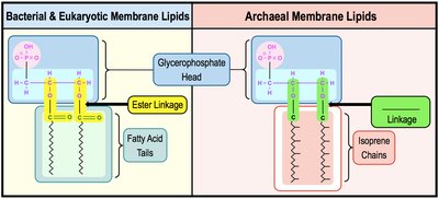
Bacterial & Eukaryotic Cell Membranes
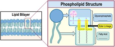
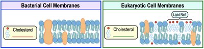
Bacterial and eukaryotic membranes are composed of phospholipids with a glycerophosphate head and fatty acid tails, connected by an ester linkage. Eukaryotic membranes contain cholesterol, which regulates membrane fluidity and forms lipid rafts.
Phospholipids: Amphipathic molecules with hydrophilic heads and hydrophobic tails.
Ester Linkage: Connects the head group to fatty acid tails in bacteria and eukaryotes.
Cholesterol: Present only in eukaryotic membranes, increasing rigidity.
Archaeal Cell Membranes
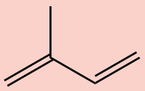
Archaeal membranes are distinct from bacterial and eukaryotic membranes in two major ways: their hydrophobic tails are composed of repeating isoprene units (not fatty acids), and the linkage between the head and tail is an ether linkage, which is more resistant to heat and chemical damage.
Isoprene Chains: 5-carbon hydrocarbon units form the hydrophobic tails.
Ether Linkage: Connects the glycerophosphate head to isoprene tails, providing stability in extreme environments.
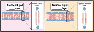
Types of Archaeal Membrane Lipids
Archaeal membrane lipids can form either bilayers or monolayers, depending on the lipid type. Monolayers, formed by diglycerol tetraether lipids, are especially common in thermophilic archaea, providing increased rigidity at high temperatures.
Bilayers: Two hydrocarbons attached to a glycerol head group (glycerol diether lipids).
Monolayers: One long hydrocarbon connects two head groups (diglycerol tetraether lipids).
Membrane Proteins
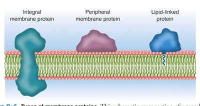
Types of Membrane Proteins
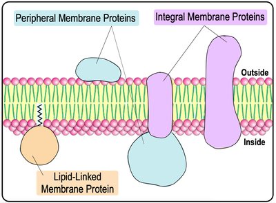
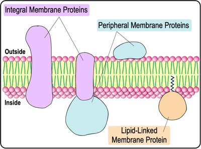
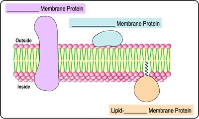
Membrane-associated proteins are classified into three main types:
Integral Proteins: Noncovalently integrated, usually spanning the entire lipid bilayer.
Peripheral Proteins: Located on the periphery of the bilayer.
Lipid-Anchored Proteins: Covalently attached to lipid groups within the bilayer.
Functions of Membrane Proteins
Membrane proteins perform a wide variety of functions, including:
Recognition: Markers for cell identification.
Anchorage: Attachment to the extracellular matrix (ECM) and cytoskeleton.
Transduction: Receptors for signal transduction pathways.
Transport: Molecular transport across the membrane.
Linkage: Connecting cells via protein linkages.
Enzymatic Activity: Catalyzing various biochemical reactions.
Concentration Gradients & Diffusion
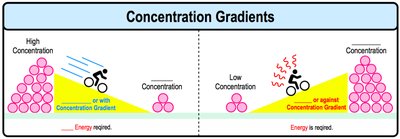
Concentration Gradients
A concentration gradient is the difference in the concentration of a substance between two areas. Molecules naturally move down their concentration gradient (from high to low concentration), a process that does not require energy.
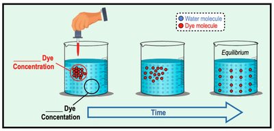
Diffusion
Diffusion is the movement of molecules from an area of higher concentration to an area of lower concentration. This process is fundamental to the movement of substances across cell membranes.
Membrane Transport
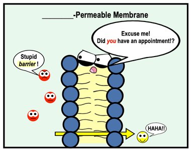
Selective Permeability
Biological membranes are selectively permeable, meaning they regulate what substances can cross. Small, uncharged, and nonpolar molecules can freely diffuse, while large, charged, or polar molecules require assistance.

Types of Membrane Transport
Transport across membranes can be classified as passive or active, and as molecular or bulk transport:
Passive Transport: No energy required; moves substances down their concentration gradient (e.g., diffusion, osmosis, facilitated diffusion).
Active Transport: Requires energy (usually ATP); moves substances against their concentration gradient.
Bulk Transport: Movement of large molecules via endocytosis or exocytosis.
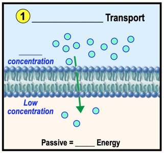
Passive Transport
Passive transport includes simple diffusion, facilitated diffusion, and osmosis. It does not require cellular energy and always moves substances from high to low concentration.
*Additional info: The above content covers the structure and function of microbial cell envelopes and membranes, including the molecular basis of membrane transport, as outlined in Chapter 2 of a standard microbiology curriculum. The images included directly reinforce the explanations of membrane structure, protein types, and transport mechanisms.*
