 Back
BackMicrobial Cell Structure and Function: Cell Morphology, Membranes, and Cell Walls
Study Guide - Smart Notes
 Tailored notes based on your materials, expanded with key definitions, examples, and context.
Cell Morphology and Arrangements
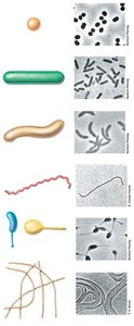
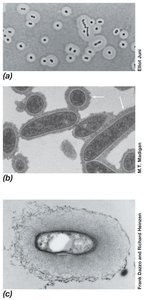
Major Cell Morphologies
Microorganisms exhibit a variety of cell shapes, known as morphologies. These shapes are important for classification and can influence microbial function and ecology.
Coccus (pl. cocci): Spherical or ovoid cells.
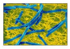
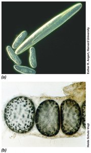
Rod (bacillus): Cylindrical, elongated cells.
Spirillum: Spiral-shaped cells.
Spirochete: Flexible, tightly coiled cells.
Appendaged bacteria: Cells with stalks or hyphae.
Filamentous bacteria: Cells forming long, thread-like chains.
Some bacteria display unusual shapes, such as stalked or budding forms, and many variations exist within these basic types.
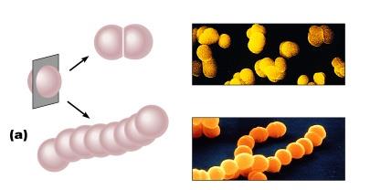
Cell Arrangements
Bacterial cells can be found in characteristic arrangements, which result from patterns of cell division and how cells remain attached after division:
Pairs: Diplococci (pairs of cocci), diplobacilli (pairs of rods)
Chains: Streptococci (chains of cocci), streptobacilli (chains of rods)
Clusters: Staphylococci (grape-like clusters of cocci)
Cell Size
The size of prokaryotic cells ranges from 0.2 µm to over 700 µm in diameter, with most rod-shaped bacteria between 0.5 and 4.0 µm wide and less than 15 µm long. Eukaryotic cells are generally larger, ranging from 10 to over 200 µm in diameter. Some bacteria, such as Epulopiscium fishelsoni and Thiomargarita namibiensis, are notable for their exceptionally large size.
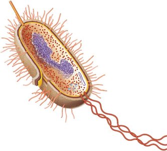
Cell Structure: Prokaryotic Cell Anatomy
General Structure of a Prokaryotic Cell
Prokaryotic cells have a relatively simple internal structure but possess specialized features for survival and adaptation:
Capsule: Protective outer layer, often polysaccharide.
Cell wall: Provides structural support and shape.
Plasma membrane: Selective barrier for transport and energy generation.
Fimbriae and pili: Surface structures for attachment and genetic exchange.
Cytoplasm: Gel-like matrix containing ribosomes, DNA (nucleoid), and inclusions.
Flagella: Motility structures.
Plasmids: Small, extrachromosomal DNA molecules.
Membrane Structure and Function
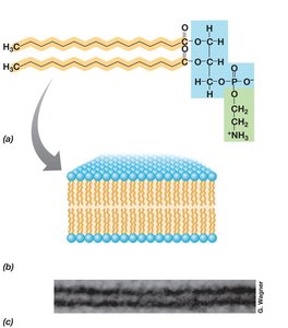
Cytoplasmic Membrane
The cytoplasmic membrane is a thin, flexible barrier that surrounds the cell, separating the cytoplasm from the external environment. It is a highly selective permeability barrier, allowing the cell to concentrate specific metabolites and excrete waste products.
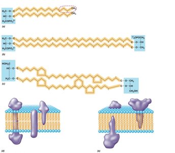
Phospholipid Bilayer
The general structure of the cytoplasmic membrane is a phospholipid bilayer composed of hydrophobic (fatty acid) tails facing inward and hydrophilic (glycerol-phosphate) heads facing outward. This arrangement creates a semi-permeable barrier.
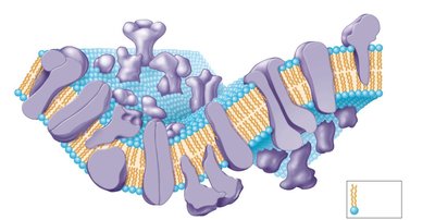
Membrane Proteins
Membrane proteins are essential for various cellular functions:
Integral membrane proteins: Firmly embedded within the membrane, often spanning the bilayer.
Peripheral membrane proteins: Loosely attached to the membrane surface.
Proteins on the outer surface interact with substrates or process large molecules for transport, while those on the inner surface are involved in energy-yielding reactions.
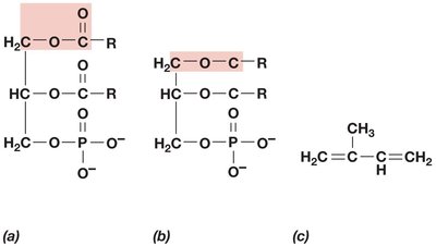
Archaeal Membranes
Archaeal membranes differ from those of Bacteria and Eukarya:
Contain ether linkages (instead of ester) in their phospholipids.
Lack fatty acids; instead, they have isoprene-based chains.
Major lipids include glycerol diethers and tetraethers, which can form monolayers, bilayers, or mixtures.
Membrane Functions
The cytoplasmic membrane serves three primary functions:
Permeability barrier: Prevents leakage and controls the entry/exit of substances.
Protein anchor: Holds proteins involved in transport, bioenergetics, and chemotaxis.
Energy conservation: Site of generation and dissipation of the proton motive force, essential for ATP synthesis.
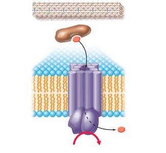
Nutrient Transport Systems
Major Classes of Transport Systems
Prokaryotes use three main types of transport systems to import nutrients:
Simple transport: Driven by the proton motive force.
Group translocation: Chemical modification of the transported substance, driven by phosphoenolpyruvate (PEP).
ATP-Binding Cassette (ABC) system: Uses periplasmic binding proteins and ATP hydrolysis for transport.
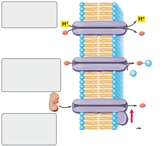
Group Translocation Example
Group translocation, such as the phosphotransferase system, involves the transfer of a phosphate group to glucose as it is transported into the cell, using energy from PEP.
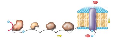
ABC Transporters
ABC transporters are the largest family of energy-driven transport systems, utilizing ATP hydrolysis to move substances across the membrane. They often involve a periplasmic binding protein, a membrane-spanning transporter, and an ATP-hydrolyzing protein.
Cell Wall Structure and Function
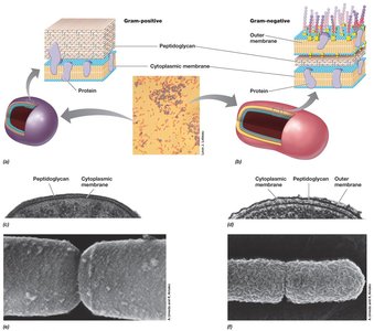
Gram-Positive vs. Gram-Negative Cell Walls
Bacteria are classified as gram-positive or gram-negative based on their cell wall structure and Gram stain reaction:
Gram-positive: Thick peptidoglycan layer, teichoic acids, no outer membrane, high susceptibility to lysozyme and penicillin.
Gram-negative: Thin peptidoglycan layer, outer membrane (LPS), periplasmic space, high susceptibility to streptomycin and tetracycline.
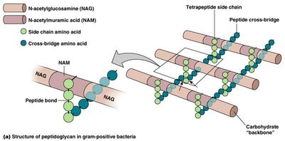
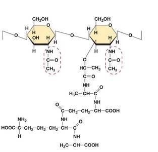
Peptidoglycan Structure
Peptidoglycan (murein) is a rigid polysaccharide layer that provides strength and resists osmotic pressure. It is composed of repeating units of N-acetylglucosamine (NAG) and N-acetylmuramic acid (NAM), cross-linked by short peptides.
The glycan backbone is the same in all bacteria.
Tetrapeptide side chains and peptide cross-bridges provide rigidity; the composition of cross-bridges varies among species.
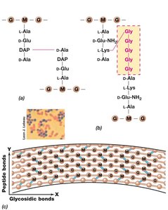
Gram-Positive Cell Wall Features
Gram-positive cell walls can contain up to 90% peptidoglycan and often have teichoic acids (acidic polysaccharides) embedded in the wall. Lipoteichoic acids are teichoic acids covalently bound to membrane lipids.
Prokaryotes Lacking Cell Walls
Some prokaryotes, such as Mycoplasma (bacteria) and Thermoplasma (archaea), lack cell walls. They survive by having sterol-like molecules in their membranes or by living in osmotically protected environments.
Outer Membrane and Lipopolysaccharide (LPS)
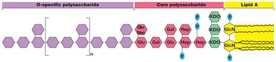
LPS Structure and Function
The outer membrane of gram-negative bacteria is rich in lipopolysaccharide (LPS), which consists of:
Lipid A: Toxic component (endotoxin)
Core polysaccharide: Contains ketodeoxyoctonate (KDO) and various sugars
O-polysaccharide: Antigenic, variable among species
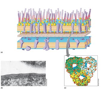
Periplasm and Porins
The periplasm is the space between the cytoplasmic and outer membranes, containing a gel-like matrix with many proteins, including those involved in nutrient transport (e.g., ABC transporters). Porins are protein channels in the outer membrane that allow passage of small hydrophilic molecules.
Gram Stain Mechanism
The structural differences in cell walls explain the Gram stain reaction:
In gram-positive cells, alcohol dehydrates the thick peptidoglycan, trapping the crystal violet-iodine complex.
In gram-negative cells, alcohol dissolves the outer membrane and leaves holes in the thin peptidoglycan, allowing the dye to wash out.
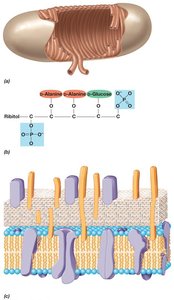
Archaeal Cell Walls
Pseudomurein and S-Layers
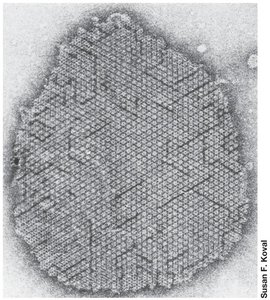
Archaeal cell walls lack peptidoglycan and typically lack an outer membrane. Some contain pseudomurein, a polysaccharide similar to peptidoglycan but composed of N-acetylglucosamine and N-acetyltalosaminuronic acid. The most common cell wall type among Archaea is the S-layer, a paracrystalline surface layer made of protein or glycoprotein, providing structural support and resistance to osmotic pressure.
Cell Surface Structures
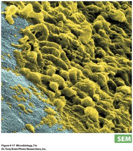
Glycocalyx: Capsules and Slime Layers
The glycocalyx is a polysaccharide layer outside the cell wall, which can form a capsule (highly organized) or a slime layer (less organized). Functions include:
Attachment to surfaces and biofilm formation
Protection against phagocytosis (increased virulence)
Resistance to desiccation
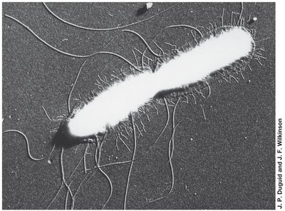
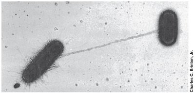
Fimbriae and Pili
Fimbriae: Short, filamentous protein structures that enable cells to adhere to surfaces or form pellicles; can contribute to virulence.
Pili: Longer than fimbriae, involved in surface attachment, genetic exchange (conjugation), and motility (type IV pili).
Cell Inclusions
Storage Granules and Magnetosomes
Bacteria often store nutrients or minerals in specialized inclusions:
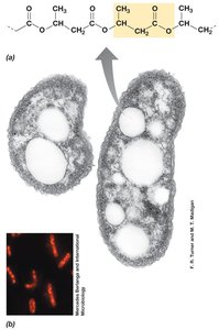
Poly-β-hydroxybutyric acid (PHB) and polyhydroxyalkanoate (PHA): Carbon storage polymers.
Glycogen: Glucose polymer for energy storage.
Polyphosphates: Inorganic phosphate storage.
Sulfur globules: Elemental sulfur storage.
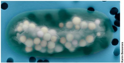
Carbonate minerals: Biomineralization of barium, strontium, and magnesium (e.g., benstonite in Gleomargarita).
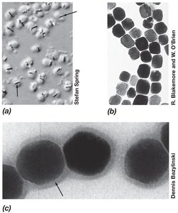
Magnetosomes: Magnetic storage inclusions for orientation in magnetic fields.
 *Additional info: Gas vesicles and endospores, as well as flagella and chemotaxis, are also important cell structures and functions but were not included in the provided images. For a complete study, students should review these topics as well.*
