 Back
Microbial Cell Structure and Function: Microscopy, Cell Walls, and Membrane Transport
Study Guide - Smart Notes
 Tailored notes based on your materials, expanded with key definitions, examples, and context.
Principles of Light Microscopy
Compound Microscopes and Their Function
Light microscopy is a foundational tool in microbiology, allowing visualization of microbial cells and structures. The compound microscope uses two sets of lenses—objective and ocular—to magnify specimens.
Magnification: The ability to make an object appear larger. Total magnification is the product of the objective and ocular lens magnifications.
Resolution: The ability to distinguish two adjacent objects as separate entities. It is determined by the wavelength of light and the numerical aperture of the lens, with a typical limit of about 0.2 μm for light microscopes.
Key Components: Ocular lenses, objective lens, stage, condenser, focusing knobs, and light source.
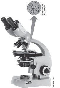
Example: Compound microscopes are used to observe stained bacterial cells, such as in the Gram stain procedure.
Gram Stain: Differential Staining Technique
Principle and Procedure
The Gram stain is a differential staining method that separates bacteria into two groups based on cell wall structure: gram-positive and gram-negative.
Gram-positive bacteria: Retain crystal violet stain and appear purple.
Gram-negative bacteria: Do not retain crystal violet after decolorization and appear red or pink after counterstaining with safranin.
Procedure:
Flood heat-fixed smear with crystal violet.
Add iodine solution (forms an insoluble complex with crystal violet).
Decolorize with alcohol.
Counterstain with safranin.
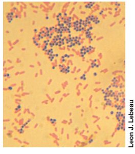
Example: In a Gram-stained field, cocci (spherical) may appear purple (G+) and rods (bacilli) may appear pink (G-).
Fluorescence Microscopy
Principle and Applications
Fluorescence microscopy is used to visualize specimens that emit light of one color when illuminated with another. This can occur naturally (autofluorescence) or after staining with fluorescent dyes such as DAPI.
Mechanism: Fluorophores absorb light at a specific wavelength and emit it at a longer wavelength, allowing for specific labeling and detection of cellular components.
Applications: Identification of specific cell types, structures, or molecules within cells.
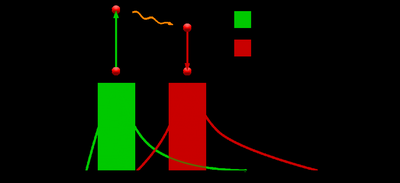
Example: Fluorescent dyes can be used to stain DNA or proteins, making them visible under a fluorescence microscope.
Imaging Cells in Three Dimensions: Confocal Scanning Laser Microscopy (CSLM)
Principle and Use
CSLM uses a laser and a computer to generate three-dimensional images by focusing on single layers of a specimen. Multiple layers are compiled to reconstruct the 3D structure.
Advantage: Provides high-resolution, three-dimensional images of cells and biofilms.
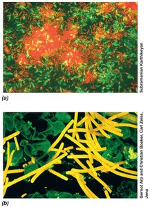
Example: CSLM is used to study the architecture of microbial communities in biofilms.
Probing Cell Structure: Electron Microscopy
Transmission and Scanning Electron Microscopes
Electron microscopes use electrons instead of photons, allowing much higher resolution (down to 0.2 nm).
Transmission Electron Microscopy (TEM): Electrons pass through thin specimens, revealing internal structures.
Scanning Electron Microscopy (SEM): Electrons scan the surface, producing detailed images of cell surfaces.
Specimen Preparation: Samples must be thin and often stained with heavy metals.
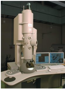
Example: TEM is used to visualize the internal organization of bacterial cells, while SEM reveals surface morphology.
Cell Morphology
Shapes and Arrangements of Microbial Cells
Microbial cells exhibit a variety of shapes, which can be important for identification but do not necessarily predict physiology or ecology.
Coccus: Spherical or ovoid.
Rod (Bacillus): Cylindrical.
Spirillum: Spiral-shaped.
Other forms: Spirochetes, appendaged, and filamentous bacteria.
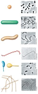
Example: Staphylococcus aureus (coccus), Escherichia coli (rod), and Spirillum volutans (spirillum).
Cell Size and the Significance of Being Small
Surface-to-Volume Ratio and Growth
Cell size affects nutrient uptake and growth rates. Small cells have a higher surface-to-volume ratio, supporting faster nutrient exchange and growth.
Typical sizes: Prokaryotes: 0.2 μm to >700 μm; Eukaryotes: 10 to >200 μm.
Advantages of small size: Greater nutrient exchange per unit volume, faster growth rates.
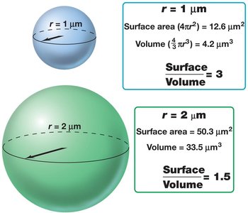
Example: Bacteria such as Thiomargarita namibiensis are unusually large, but most bacteria are much smaller to maximize nutrient uptake.
Bacterial Membrane Structure
Phospholipid Bilayer and Membrane Proteins
The cytoplasmic membrane is a vital, selectively permeable barrier composed of a phospholipid bilayer with embedded proteins.
Phospholipids: Contain hydrophobic fatty acid tails and hydrophilic heads.
Membrane proteins: Integral (embedded) and peripheral (loosely attached).
Stabilization: Hydrogen bonds, hydrophobic interactions, and ionic bonds with Mg2+ and Ca2+.
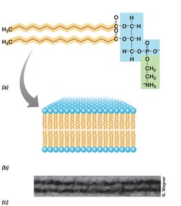
Example: The fluid mosaic model describes the dynamic nature of the membrane, with proteins moving within the lipid bilayer.
Archaeal Membrane Structure
Unique Features of Archaeal Membranes
Archaeal membranes differ from bacterial and eukaryotic membranes in their chemical composition and structure.
Ether linkages (instead of ester) connect glycerol to hydrophobic side chains.
Isoprenoid chains (phytanyl or biphytanyl) replace fatty acids.
Lipid monolayers (in some Archaea) provide extra stability in extreme environments.
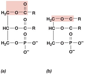
Example: Thermophilic Archaea often have lipid monolayers for increased membrane stability at high temperatures.
Membrane Function and Transport
Roles of the Cytoplasmic Membrane
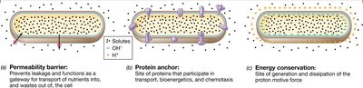
The cytoplasmic membrane serves as a permeability barrier, protein anchor, and site of energy conservation (proton motive force generation).
Permeability barrier: Prevents leakage and controls entry/exit of substances.
Protein anchor: Holds transport and bioenergetic proteins.
Energy conservation: Maintains proton gradients for ATP synthesis.
Transport Mechanisms
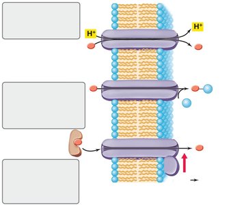
Microbial cells use various transport systems to move nutrients across the membrane, often against concentration gradients.
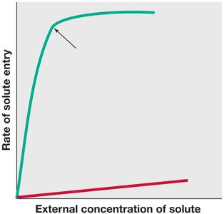
Simple diffusion: Passive movement down a concentration gradient.
Carrier-mediated transport: Involves specific transport proteins and shows saturation kinetics.
Types of Transport Events
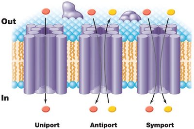
Uniport: One substance moves in one direction.
Symport: Two substances move in the same direction together.
Antiport: Two substances move in opposite directions.
Major Classes of Transport Systems
Simple transport: Driven by the proton motive force.
Group translocation: Chemical modification of the transported substance (e.g., phosphotransferase system).
ABC transporters: Use ATP hydrolysis and substrate-binding proteins for high specificity.
Cell Walls of Bacteria and Archaea
Peptidoglycan Structure and Gram Stain Differences
Bacterial cell walls are composed of peptidoglycan, a unique polymer that provides structural strength. Gram-positive and gram-negative bacteria differ in cell wall architecture.
Gram-positive: Thick peptidoglycan layer, often with teichoic acids.
Gram-negative: Thin peptidoglycan layer and an outer membrane containing lipopolysaccharide (LPS).
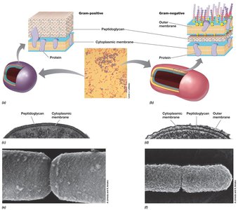
Example: Staphylococcus aureus is Gram-positive; Escherichia coli is Gram-negative.
Peptidoglycan Chemical Structure
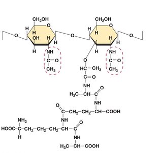
Peptidoglycan consists of repeating units of N-acetylglucosamine (G) and N-acetylmuramic acid (M), cross-linked by peptides containing unusual amino acids.
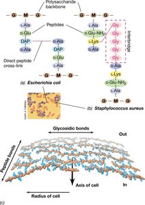
Cross-linking: Varies between Gram-positive and Gram-negative bacteria.
Antibiotic target: Peptidoglycan synthesis is inhibited by several antibiotics (e.g., penicillin).
Cross-linking in Peptidoglycan
The glycan chains are cross-linked by peptides, which differ in composition and linkage between Gram-positive and Gram-negative bacteria.
Summary Table: Comparison of Cell Envelope Structures
Feature | Gram-Positive Bacteria | Gram-Negative Bacteria | Archaea |
|---|---|---|---|
Peptidoglycan | Thick, up to 90% | Thin, ~10% | Absent (pseudomurein or S-layer) |
Outer Membrane | Absent | Present (LPS layer) | Absent |
Teichoic Acids | Present | Absent | Absent |
Lipid Composition | Ester-linked fatty acids | Ester-linked fatty acids | Ether-linked isoprenoids |
Additional info: This table summarizes the main differences in cell envelope structure among Gram-positive, Gram-negative bacteria, and Archaea, which are critical for understanding staining reactions, antibiotic susceptibility, and environmental adaptations.
