 Back
Microbial Cell Structure & Function: Prokaryotic and Eukaryotic Cells
Study Guide - Smart Notes
 Tailored notes based on your materials, expanded with key definitions, examples, and context.
Microbial Cell Structure & Function
Structure & Composition of Microbial Cells
Microbial cells exhibit diverse structural features that are essential for their survival, growth, and interaction with their environment. The cell structure determines the cell's function, pathogenicity, and response to external stimuli.
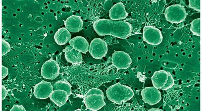
Cell Morphology: Microbes display a variety of shapes, including cocci (spherical), rods (cylindrical), spirilla (spiral), spirochetes, appendaged, and filamentous forms.
Cell Wall: Provides structural support, protection from osmotic stress, and is a target for immune responses and antibiotics.
Plasma Membrane: Acts as a selective barrier and is involved in energy transformation and transport.
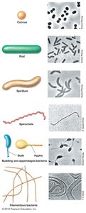
Prokaryotic Cell Morphology
Prokaryotes come in a variety of shapes, which are fundamental to their identification and classification.
Coccus: Spherical or ovoid shape.
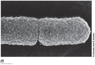
Rod: Cylindrical shape.
Spirillum: Spiral shape.
Unusual shapes: Spirochetes, appendaged bacteria, filamentous bacteria.
Morphological diversity: Many species fall into these basic categories, but extensions and variations exist.
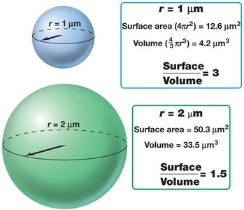
The Advantage of Being Small
Small microbial cells have a high surface area-to-volume ratio, which is advantageous for nutrient uptake and rapid growth.
Surface Area: Total area occupied by the cell's surface.
Volume: Space enclosed by the cell.
High Surface Area-to-Volume Ratio: Facilitates efficient nutrient exchange and supports faster growth and replication.
Evolutionary Implications: Higher replication rates increase evolutionary change and diversity.
Size Limitations: Cells smaller than 1 µm may lack essential macromolecules.
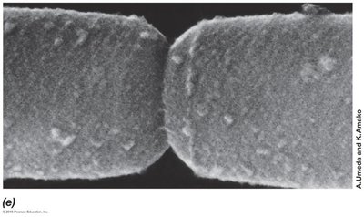
The Cell Wall: Protection and Interaction
The cell wall is a critical structure for microbial cells, providing shape, protection, and interaction with the environment.
Biological Significance: Maintains cell shape and prevents lysis under osmotic stress.
Clinical Significance: Cell wall molecules are antigenic and recognized by the host immune system; cell wall is a target for antibiotics (e.g., penicillin) and lysozyme.
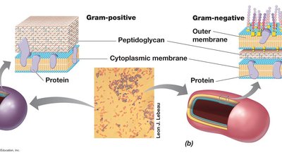
Gram Positive & Gram Negative Cell Walls
Bacterial cell walls are classified as Gram-positive or Gram-negative based on their structure and staining properties.
Gram-positive: Thick peptidoglycan layer, contains teichoic acids, net negative charge, sensitive to penicillin.
Gram-negative: Thin peptidoglycan layer, outer membrane with lipopolysaccharide (LPS), provides protection from antibodies and toxins.
Peptidoglycan: Macromolecule unique to bacteria, provides rigidity.
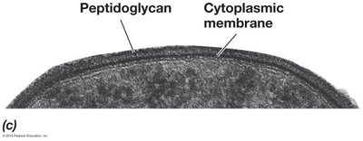
Composition of the Gram-Positive Cell Wall
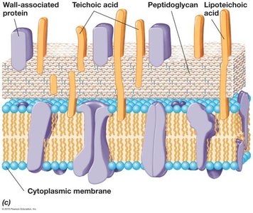
Gram-positive cell walls are characterized by a thick peptidoglycan layer and the presence of teichoic acids.
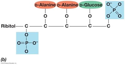
Teichoic Acids: Polymers of ribitol or glycerol linked by phosphate bonds; contribute to negative charge.
Peptidoglycan: Provides rigidity and is the target of penicillin.
Characteristics of the Peptidoglycan Layer
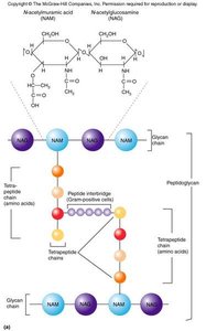
Peptidoglycan is a complex polymer that forms the backbone of bacterial cell walls.
Glycan Chain: Composed of N-acetylglucosamine (NAG) and N-acetylmuramic acid (NAM).
Tetrapeptide Chain: Four amino acids cross-link glycan chains; includes D-glutamic acid, D-alanine, and diaminopimelic acid.
Composition of the Gram-Negative Cell Wall
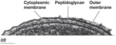
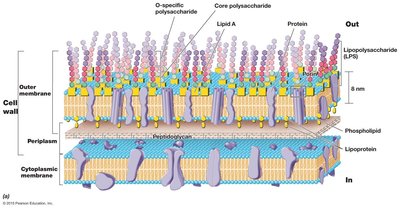
Gram-negative cell walls have a thin peptidoglycan layer and an outer membrane containing LPS.
Outer Membrane: Lipid bilayer acts as a protective barrier.
LPS: Outer leaflet of the membrane, provides structural integrity and protection.
Clinical Properties: Protects from antibodies and toxins.
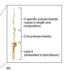
Lipopolysaccharide (LPS) Structure and Function
LPS is a major component of the Gram-negative outer membrane and is important for pathogenicity.
Lipid A: Embedded in the lipid bilayer, toxic to humans (endotoxin).
Core Polysaccharide: Connects Lipid A to O side chain.
O Side Chain: Varies between species, important for immune recognition.
Gram Staining: Why Do Bacterial Cells Stain Differentially?
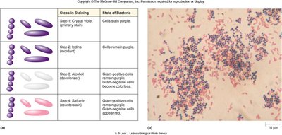
Gram staining differentiates bacteria based on cell wall structure.
Crystal Violet-Iodine: Stains intracellular contents.
Alcohol-Acetone: Alters peptidoglycan layer; traps dye in Gram-positive, removes dye in Gram-negative.
Clinical Relevance: Gram-positive cells retain purple stain; Gram-negative cells appear red after counterstain.
Bacteria Lacking Cell Walls
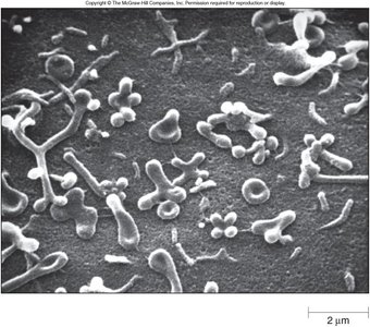
Some bacteria, such as Mycoplasma, lack rigid cell walls and are resistant to certain antibiotics.
Clinical Relevance: Resistant to penicillin and lysozyme.
Unique Plasma Membrane: Compensates for lack of cell wall.
Surface Layers External to the Cell Wall: Capsules & Slime Layers
Many bacteria produce external layers called capsules and slime layers, collectively known as glycocalyx.
Composition: Predominantly carbohydrates.
Biological Significance: Adherence to surfaces (e.g., dental plaque), evasion of immune defenses.
The Cytoplasmic (Plasma) Membrane: Barrier
The plasma membrane is a selective barrier that separates the cell's internal environment from the external environment.
Structure: Lipid bilayer with embedded proteins.
Function: Selective transport, energy transformation, fluid mosaic model.
Membrane Transport Mechanisms
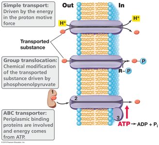
Microbial cells utilize various mechanisms to transport substances across the plasma membrane.
Simple Transport: Driven by proton motive force.
Group Translocation: Chemical modification of transported substance.
ABC Transporter: Uses ATP for energy, involves periplasmic binding proteins.
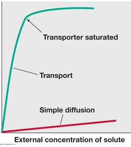
Transport Saturation: Transporters can become saturated, limiting rate of transport.
The Flagella: Movement
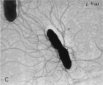
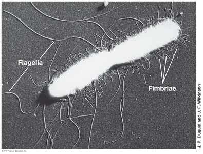
Flagella are appendages used for motility, allowing bacteria to move toward or away from environmental cues.
Types: Peritrichous (distributed), polar (single at one end), lophotrichous (multiple at one end).
Movement: Directed by taxis (chemotaxis, phototaxis, aerotaxis, magnetotaxis, thermotaxis, hydrotaxis).
The Pilus: Adhesion & DNA Transfer
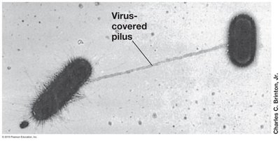
Pili are structures used for adhesion and genetic exchange (conjugation) in bacteria.
Pilus: Used for conjugation, joining two cells for DNA transfer.
Fimbriae: Mediate adherence to surfaces, coated with adhesive proteins.
The Bacterial Chromosome: Information
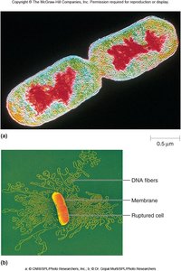
Bacterial genetic information is stored in a single, circular chromosome and may be supplemented by plasmids.
Chromosome: Circular, double-stranded DNA in nucleoid space.
Plasmid: Small, circular DNA, extrachromosomal, encodes genes for survival (e.g., antimicrobial resistance).
The Ribosome: Synthesis
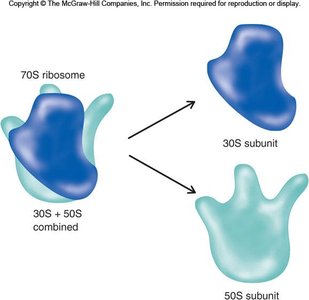
Ribosomes are the site of protein synthesis in microbial cells.
Structure: 70S ribosome composed of 30S and 50S subunits (rRNA and protein).
Sedimentation Rate: S value reflects size and density, not additive.
Storage Vesicles (Granules/Gas Vesicles)
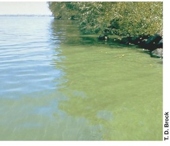
Storage granules and gas vesicles are specialized structures for storing molecules and regulating buoyancy.
Storage Granules: Store organic and inorganic molecules (e.g., phosphate).
Gas Vesicles: Used by aquatic bacteria to float at optimal depth for nutrient acquisition.
The Endospore: Survival
Endospores are dormant, highly durable cell types formed by certain bacteria for survival under harsh conditions.
Formation: Produced by Bacillus and Clostridium species.
Durability: Resistant to desiccation, heat, UV, and chemicals; can survive for decades.
Clinical Relevance: Some endospore-forming species cause disease (e.g., botulism, tetanus, anthrax).
Eukaryotic Cell Structure
Eukaryotic microbes (algae, fungi, protozoa) possess membrane-bound organelles and complex internal structures.
Organelles: Nucleus, mitochondria, chloroplast, endoplasmic reticulum, Golgi apparatus.
Cytoskeleton: Provides structural support and facilitates movement.
Cell Wall: Present in fungi and algae, different composition from bacterial cell walls.
Review: Prokaryotes & Eukaryotes
Prokaryotes and eukaryotes differ fundamentally in cell structure, complexity, and organelle presence.
Prokaryotes: Lack membrane-bound organelles, have a single circular chromosome, cell wall composed of peptidoglycan.
Eukaryotes: Possess membrane-bound organelles, multiple linear chromosomes, cell wall (if present) composed of different molecules.
