 Back
BackMicrobial Cell Structure and Function: Study Guide
Study Guide - Smart Notes
 Tailored notes based on your materials, expanded with key definitions, examples, and context.
Microbial Cell Structure and Function
Overview of Microbial Cell Structures
Microbial cells possess a variety of structural components that enable them to interact with their environment, maintain integrity, and perform essential functions. The cell envelope is a key feature, providing protection and mediating exchanges with the surroundings.
Cytoplasmic (Cell) Membrane: Selective permeability, energy metabolism
Cell Wall: Maintains shape, rigidity, prevents lysis
Outer Membrane (Gram-negative only): Protection, virulence
Capsule: Virulence, immune evasion
Fimbriae: Attachment
Pili: DNA transfer (conjugation)
Flagella: Motility
Inclusions: Nutrient and energy storage
Endospores: Survival under harsh conditions
The Cell Envelope
The cell envelope is a layered system surrounding the cytoplasm, crucial for protection and environmental interaction.
Cytoplasmic membrane: Present in all cells
Cell wall: Most bacteria
Outer membrane: Gram-negative only
S-layer: Some Bacteria & Archaea
Cytoplasmic (Cell) Membrane
The cytoplasmic membrane is a phospholipid bilayer, typically 8–10 nm thick, with embedded proteins. It is essential for selective permeability and energy metabolism.
Structure: Hydrophobic fatty acid tails inward, hydrophilic phosphate heads outward
Functions: Selective permeability, nutrient uptake, waste removal, energy metabolism, protein anchoring
Membrane Proteins: Integral (transmembrane) and peripheral (loosely attached)
Membrane Differences Across Domains
Bacteria and Eukarya have ester-linked fatty acids, while Archaea possess ether-linked isoprenoid chains, which can form lipid monolayers for extreme stability.
Bacteria & Eukarya: Ester linkages, fatty acid chains
Archaea: Ether linkages, isoprenoid chains, lipid monolayers
Cell Wall
The cell wall prevents osmotic lysis and maintains cell shape. In bacteria, it is composed of peptidoglycan, a polymer of N-acetylglucosamine (NAG) and N-acetylmuramic acid (NAM) linked by β-1,4 glycosidic bonds and cross-linked peptides.
Peptidoglycan: Unique to bacteria
Absent in Archaea & Eukarya

Gram-Positive vs Gram-Negative Bacteria
Bacteria are classified based on their cell wall structure, which affects their staining properties and susceptibility to antibiotics.
Gram-Positive: Thick peptidoglycan, teichoic acids, no outer membrane (e.g., Staphylococcus)
Gram-Negative: Thin peptidoglycan, outer membrane with LPS, periplasmic space (e.g., E. coli)
Lysozyme & Antibiotics
Lysozyme and antibiotics target the bacterial cell wall, leading to cell lysis.
Lysozyme: Breaks β-1,4 bonds in peptidoglycan
Penicillin: Blocks peptide cross-linking
Archaeal Cell Walls
Archaea lack peptidoglycan and often possess S-layers. Methanogens have pseudomurein, which is resistant to lysozyme and penicillin due to β-1,3 bonds and all L-amino acids.
S-layers: Protein shell
Pseudomurein: Peptidoglycan-like polymer
Gram-Negative Outer Membrane (LPS)
The outer membrane of Gram-negative bacteria contains lipopolysaccharide (LPS), which is important for virulence and immune response.
LPS Components: O-polysaccharide (antigenic), core polysaccharide, lipid A (endotoxin)
Other Features: Porins (transport), Braun lipoprotein (anchors OM), periplasm (enzymes & proteins)
S-Layers
S-layers are protein or glycoprotein lattices found in some Bacteria and Archaea, providing protection, shape, and adhesion.
Function: Protection, shape, adhesion
Cell Surface Structures
Microbial cells may possess capsules, slime layers, pili, fimbriae, and hami, which aid in attachment, biofilm formation, and immune evasion.
Capsules & Slime Layers: Polysaccharide coating, prevent phagocytosis, aid in biofilms
Pili & Fimbriae: Fimbriae (attachment), sex pili (conjugation), type IV pili (twitching motility)
Hami (Archaea): Grappling-hook structures for attachment
Cell Inclusions
Cell inclusions are storage structures for nutrients and energy, as well as specialized functions like buoyancy and magnetotaxis.
Types: PHB/PHA (carbon storage), glycogen, polyphosphate granules, sulfur granules, carbonate minerals, gas vesicles (buoyancy), magnetosomes (magnetotaxis)
Endospores
Endospores are highly resistant survival structures formed by certain Gram-positive bacteria, such as Bacillus and Clostridium.
Components: Dipicolinic acid + Ca2+, SASPs (protect DNA)
Germination Stages: Activation, germination, outgrowth
Flagella & Motility
Flagella are motility structures powered by the proton motive force. Their arrangement varies among species.
Arrangements: Polar, lophotrichous, amphitrichous, peritrichous
Energy Source: Proton motive force
Surface Motility
Microbes can move across surfaces using twitching (type IV pili + ATP) or gliding (smooth movement without flagella).
Twitching: Type IV pili + ATP
Gliding: Smooth movement
Chemotaxis & Taxis
Chemotaxis is movement toward or away from chemicals, while other taxis include responses to light, oxygen, osmotic pressure, water, and magnetic fields.
Chemotaxis: Run & tumble (e.g., E. coli)
Other Taxis: Phototaxis, aerotaxis, osmotaxis, hydrotaxis, magnetotaxis
Endosymbiotic Theory
The endosymbiotic theory explains the origin of mitochondria and chloroplasts in eukaryotic cells, based on evidence such as circular DNA, bacterial-like ribosomes, and double membranes.
Mitochondria: Derived from bacteria

Chloroplasts: Derived from cyanobacteria

Microbial Morphology and Arrangement
Bacteria exhibit diverse shapes and arrangements, which are important for identification and classification.
Cocci: Spherical (e.g., Staphylococcus)
Rods (bacilli): Elongated (e.g., E. coli)
Spirilla: Spiral-shaped
Arrangement: Single, chains, clusters, tetrads
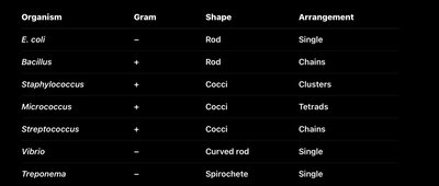
Organism | Gram | Shape | Arrangement |
|---|---|---|---|
E. coli | − | Rod | Single |
Bacillus | + | Rod | Chains |
Staphylococcus | + | Cocci | Clusters |
Micrococcus | + | Cocci | Tetrads |
Streptococcus | + | Cocci | Chains |
Vibrio | − | Curved rod | Single |
Treponema | − | Spirochete | Single |
Prokaryotic vs Eukaryotic Cells
Prokaryotes (bacteria, archaea) lack a nucleus and organelles, while eukaryotes (animals, plants, fungi) possess these structures.
Prokaryotes: Bacteria, Archaea
Eukaryotes: Animals, plants, fungi
Reproduction
Microbial reproduction varies by domain and group.
Bacteria: Binary fission
Fungi: Spores/budding
Plants: Sexual & asexual
Animals: Sexual
Endospores vs Fungal Spores
Endospores are survival structures, while fungal spores are reproductive.
Endospores: Survival
Fungal spores: Reproduction
Phototaxis
Phototaxis is the movement of microorganisms toward or away from light, an important behavior for photosynthetic microbes.
Positive phototaxis: Toward light
Negative phototaxis: Away from light
Do Bacteria Have Cilia?
Bacteria do not possess cilia; cilia are exclusive to eukaryotic cells.

Key Equations and Concepts
Peptidoglycan Structure:
Binary Fission:
Proton Motive Force:
Summary Table: Gram-Positive vs Gram-Negative
Feature | Gram-Positive | Gram-Negative |
|---|---|---|
Peptidoglycan | Thick | Thin |
Teichoic acids | Present | Absent |
Outer membrane | Absent | Present |
LPS | Absent | Present |
Periplasm | Absent | Present |
Additional info: Academic context was added to expand brief points and clarify structural and functional differences among microbial cell types, as well as to provide examples and key equations for exam preparation.
