 Back
BackMicrobial Cell Structure and Function: Study Notes for Microbiology Students
Study Guide - Smart Notes
 Tailored notes based on your materials, expanded with key definitions, examples, and context.
Structure and Function of Bacterial Cells
Exploring the Microbial Cell
The microbial cell is the fundamental unit of life in microbiology, exhibiting a variety of structures and functions that enable survival and adaptation. Understanding cell structure is essential for appreciating microbial diversity and unity.
Microscopy is crucial for visualizing microbial cells and their components.
Microbial cultivation expands our knowledge of microbial physiology and genetics.
Microbiology helps elucidate the unity and diversity of life.
The Cell Envelope
Cytoplasmic Membrane
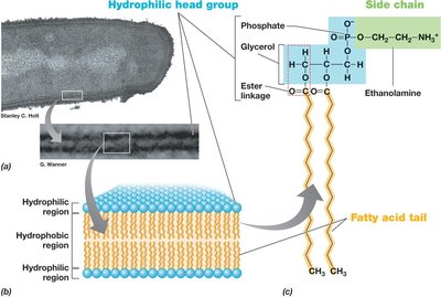
The cytoplasmic membrane is a selectively permeable barrier that separates the cytoplasm from the external environment. It is essential for nutrient transport, energy metabolism, and maintaining cellular integrity.
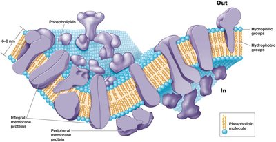
Structure: Phospholipid bilayer with embedded proteins; 8–10 nm wide.
Hydrophobic fatty acid tails face inward, while hydrophilic head gr oups face outward.
Membrane proteins: Integral, transmembrane, and peripheral proteins facilitate transport and energy processes.
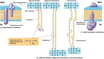
Example: The phospholipid bilayer structure is common to both Bacteria and Eukarya, but Archaea have ether linkages and isoprenoid chains.
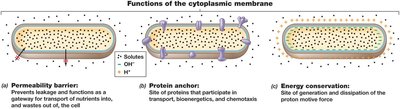
Cytoplasmic Membrane Functions
Permeability barrier: Prevents leakage and controls entry/exit of substances.
Protein anchor: Holds proteins involved in transport and energy metabolism.
Energy conservation: Generates proton motive force for ATP synthesis.
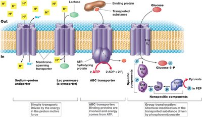
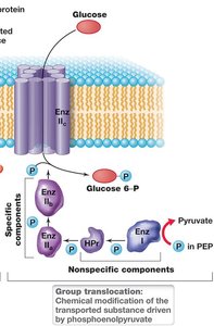
Transporting Nutrients into the Cell
Microbial cells use specialized transport systems to accumulate solutes against concentration gradients. These systems are energy-driven and essential for survival in diverse environments.
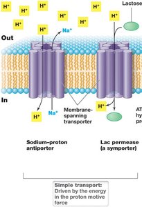
Simple transport: Uses transmembrane proteins and proton motive force.
Group translocation: Involves chemical modification of the transported substance (e.g., phosphotransferase system).
ABC transporter system: Uses ATP and substrate-binding proteins for high-affinity uptake.

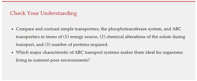
The Cell Wall
Structure and Function
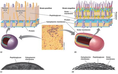
The cell wall provides structural support, resists osmotic pressure, and maintains cell shape. Most bacteria are classified as gram-positive or gram-negative based on cell wall structure.
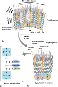
Gram-positive: Thick peptidoglycan layer, teichoic acids, no outer membrane.
Gram-negative: Thin peptidoglycan layer, outer membrane, periplasmic space.
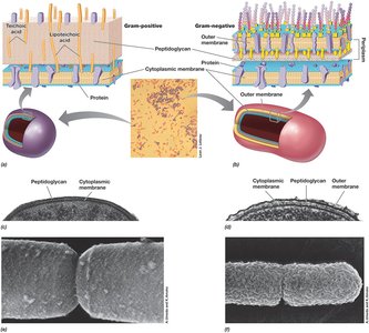
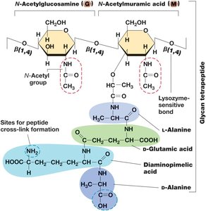
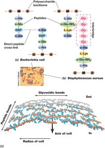
Peptidoglycan Structure
Peptidoglycan is a rigid polysaccharide layer unique to bacteria, composed of alternating N-acetylglucosamine and N-acetylmuramic acid, cross-linked by peptides.
Provides strength and resists turgor pressure.
Gram-negative: Single layer, cross-links between DAP and D-alanine.
Gram-positive: Multiple layers, peptide interbridges (e.g., five glycines in Staphylococcus aureus).
Archaeal Cell Walls
Archaea lack peptidoglycan and typically lack an outer membrane. Most have an S-layer (protein shell) or, in methanogens, a pseudomurein cell wall.
Pseudomurein: Alternating N-acetylglucosamine and N-acetyltalosaminuronic acid, β-1,3 linkages, all L-amino acids.
Resistant to lysozyme and penicillin.
LPS: The Outer Membrane
Gram-Negative Cell Envelope
The outer membrane is a second lipid bilayer external to the cell wall in gram-negative bacteria. It contains lipopolysaccharide (LPS), which is important for surface recognition, virulence, and structural strength.
LPS: Composed of lipid A (endotoxin), core polysaccharide, and O-polysaccharide.
Porins: Transmembrane proteins for solute transport.
Periplasm: Space between cytoplasmic and outer membranes, contains extracellular proteins.
Diversity of Cell Envelope Structure
S-Layers and Alternative Configurations
S-layers are paracrystalline protein or glycoprotein structures that provide strength, protection, and facilitate cell surface interactions. Some bacteria and archaea lack cell walls but have tough cytoplasmic membranes.
S-layer: Always the outermost layer if present.
Mycoplasmas: Bacteria lacking cell walls, contain sterols.
Thermoplasma: Archaea lacking cell walls.
Cell Surface Structures and Inclusions
Capsules and Slime Layers
Capsules and slime layers are sticky polysaccharide coats outside the cell envelope, aiding in attachment, biofilm formation, infectivity, and protection from desiccation.
Capsule: Tightly attached, visible with India ink.
Slime layer: Loosely attached, easily deformed.
Fimbriae, Pili, and Hami
Fimbriae and pili are protein structures that mediate attachment, biofilm formation, and genetic exchange. Hami are unique archaeal structures resembling grappling hooks.
Fimbriae: Short pili for attachment.
Pili: Longer, involved in conjugation and motility.
Hami: Barbed termini for surface attachment in Archaea.
Cell Inclusions
Cell inclusions serve as energy reserves, carbon or phosphorus storage, and have specialized functions. They are enclosed by thin protein membranes to reduce osmotic stress.
Carbon storage: Poly-β-hydroxybutyric acid (PHB), poly-β-hydroxyalkanoate (PHA), glycogen.
Polyphosphate granules: Inorganic phosphate storage.
Elemental sulfur: Accumulates in periplasmic granules.
Carbonate minerals: Biomineralization of barium, strontium, magnesium.
Gas vesicles: Confer buoyancy.
Magnetosomes: Biomineralized magnetic iron oxides for magnetotaxis.
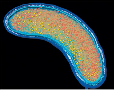
Endospores
Endospores are highly differentiated, dormant cells resistant to extreme conditions. They are survival structures formed by some gram-positive bacteria.
Formation: Triggered by nutrient limitation; involves activation, germination, and outgrowth.
Structure: Multiple layers, dipicolinic acid, small acid-soluble spore proteins (SASPs).
Table: Differences between endospores and vegetative cells.
Characteristic | Vegetative cell | Endospore |
|---|---|---|
Microscopic appearance | Nonrefractile | Refractile |
Calcium content | Low | High |
Dipicolinic acid | Absent | Present |
Enzymatic activity | High | Low |
Respiration rate | High | Low or absent |
Macromolecular synthesis | Present | Absent |
Heat resistance | Low | High |
Radiation resistance | Low | High |
Resistance to chemicals | Low | High |
Lysozyme | Sensitive | Resistant |
Water content | High, 80–90% | Low, 10–25% in core |
Small acid-soluble spore proteins | Absent | Present |
Cell Locomotion
Flagella, Archaella, and Swimming Motility
Flagella and archaella are motility structures that enable swimming in Bacteria and Archaea. They are tiny rotating machines anchored in the cell, with various arrangements and speeds.
Flagella: Long, thin appendages; arrangements include polar, tufts, lophotrichous, amphitrichous, peritrichous.
Structure: Filament (flagellin), hook, basal body (motor).
Archaella: Smaller, unrelated proteins, ATP-driven rotation.
Surface Motility
Surface motility includes twitching and gliding, which are slower than swimming and involve type IV pili or intracellular protein tracks.
Twitching: Extension and retraction of pili.
Gliding: Smooth motion along cell axis, only in Bacteria.
Chemotaxis and Other Forms of Taxis
Taxis is directed movement in response to chemical or physical stimuli. Chemotaxis, phototaxis, aerotaxis, osmotaxis, and hydrotaxis allow microbes to optimize their position for resources or avoid harm.
Run and tumble: Movement pattern in peritrichously flagellated bacteria.
Phototaxis: Response to light; scotophobotaxis is response to darkness.
Aerotaxis: Response to oxygen concentration.
Magnetotaxis: Movement along magnetic field lines.
Eukaryotic Microbial Cells
The Nucleus and Cell Division
Eukaryotic cells contain a double membrane-enclosed nucleus and other organelles. Cell division occurs via mitosis or meiosis.
Nucleus: Contains chromosomes, nucleolus (site of rRNA synthesis).
Mitosis: Produces two diploid daughter cells.
Meiosis: Produces four haploid gametes.
Mitochondria and Chloroplasts
Mitochondria and chloroplasts specialize in energy metabolism and have evolutionary roots within Bacteria. They provide ATP and are sites of respiration and photosynthesis, respectively.
Mitochondria: Respiration, oxidative phosphorylation, cristae, matrix.
Chloroplasts: Photosynthesis, thylakoids, stroma, RuBisCO enzyme.
Endosymbiotic hypothesis: Organelles descended from bacterial cells.
Other Eukaryotic Cell Structures
The cytoskeleton provides internal structural support, while the endoplasmic reticulum, Golgi complex, and lysosomes are involved in synthesis, modification, and recycling. Flagella and cilia enable motility.
Cytoskeleton: Microtubules, microfilaments, intermediate filaments.
Endoplasmic reticulum: Rough (ribosomes), smooth (lipid synthesis).
Golgi complex: Modifies ER products.
Lysosomes: Digestive enzymes, recycling.
Flagella and cilia: Motility organelles, structurally distinct from prokaryotic flagella.
Additional info: Academic context and explanations have been expanded for clarity and completeness. Only images directly relevant to the explanation have been included.
