 Back
BackMicrobial Cell Structure and Function: Study Notes
Study Guide - Smart Notes
 Tailored notes based on your materials, expanded with key definitions, examples, and context.
Microbial Cell Structure and Function
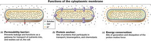
The Cytoplasmic Membrane
The cytoplasmic membrane is a fundamental structure in all microorganisms, serving as a boundary between the cell's internal environment and the external surroundings. Its primary function is selective permeability, allowing the transport of nutrients into the cell and the removal of waste products. Membrane proteins embedded within the membrane facilitate these processes and play a crucial role in energy metabolism.
Selective Permeability: The membrane acts as a barrier, controlling the entry and exit of substances.
Membrane Proteins: These proteins are involved in transport, biosynthesis, chemotaxis, and energy conservation.
Energy Metabolism: The membrane is the site of generation and dissipation of the proton motive force, essential for ATP synthesis.
Structure: The cytoplasmic membrane is composed of a phospholipid bilayer, with hydrophobic tails facing inward and hydrophilic heads facing outward.
Phospholipid Bilayer Membrane
The phospholipid bilayer is the basic structural component of the cytoplasmic membrane. It provides fluidity and flexibility, allowing the membrane to self-heal and adapt to environmental changes.
Phospholipids: Amphipathic molecules with hydrophilic heads and hydrophobic tails.
Fluid Mosaic Model: Describes the dynamic nature of the membrane, with proteins and lipids moving laterally.
Major Lipids of Archaea and the Architecture of Archaeal Membranes
Archaeal membranes differ significantly from bacterial membranes. Archaea possess unique lipids, such as ether-linked phospholipids, which provide stability under extreme conditions.
Ether Linkages: Archaeal lipids contain ether bonds, unlike the ester bonds in bacterial lipids.
Monolayer Structure: Some archaeal membranes are monolayers, enhancing resistance to heat and chemical stress.
Comparison of Bacterial and Archaeal Membranes
Bacterial and archaeal membranes exhibit distinct structural and chemical differences, reflecting their evolutionary divergence.
Bacterial Membranes: Composed of ester-linked phospholipids.
Archaeal Membranes: Composed of ether-linked lipids, often forming monolayers.
The Cell Wall
The cell wall is a rigid structure that provides shape and protection to microbial cells. It prevents cell lysis due to osmotic pressure and is a key determinant in the classification of bacteria.
Osmotic/Turgor Pressure: The cell wall withstands internal pressure, preventing rupture.
Shape and Rigidity: Maintains the structural integrity of the cell.
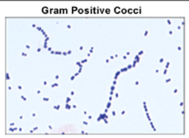
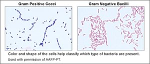
Gram Stain: Bacteria are classified as Gram-positive or Gram-negative based on cell wall structure.
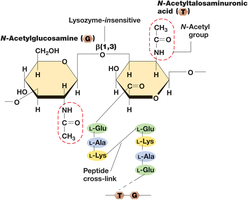
Structure of the Repeating Unit in Peptidoglycan: The Glycan Tetrapeptide
Peptidoglycan is a complex polymer forming the main component of bacterial cell walls. It consists of repeating units of N-acetylglucosamine and N-acetylmuramic acid, linked by peptide cross-bridges.
Glycan Tetrapeptide: Alternating sugar residues with attached tetrapeptide chains.
Cross-linking: Provides strength and rigidity to the cell wall.
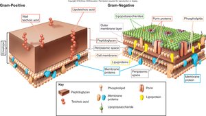
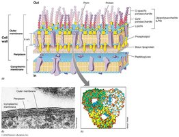
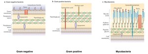
Structural Differences: Gram-Positive vs Gram-Negative Cell Walls
Gram-positive and Gram-negative bacteria differ in cell wall composition and structure, which affects their staining properties and susceptibility to antibiotics.
Gram-Positive: Thick peptidoglycan layer, teichoic acids, no outer membrane.
Gram-Negative: Thin peptidoglycan layer, outer membrane with lipopolysaccharide (LPS), periplasmic space.
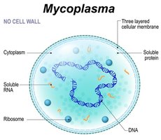
Do All Prokaryotes Have a Cell Wall?
Not all prokaryotes possess a cell wall. Some, such as Mycoplasma, lack a cell wall and rely on a robust cytoplasmic membrane for structural integrity.
Mycoplasma: No cell wall, three-layered cellular membrane.
Adaptations: Resistance to osmotic stress and certain antibiotics.
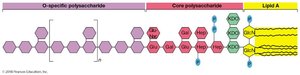
Most of Cell Wall Composed of Outer Membrane or Lipopolysaccharide (LPS) Layer
In Gram-negative bacteria, the outer membrane is a major component of the cell wall, with LPS replacing most phospholipids in the outer leaflet. LPS contains lipid A, which acts as an endotoxin.
LPS: Composed of O-specific polysaccharide, core polysaccharide, and lipid A.
Endotoxin: Lipid A is toxic and can trigger immune responses.
Alternative Configurations of the Cell Wall
Microbial cell walls exhibit diverse configurations, including variations in peptidoglycan structure and the presence of unique polymers in certain groups.
Mycobacteria: Contain mycolic acids, making the cell wall waxy and resistant to staining.
Other Variants: Some bacteria and archaea have specialized cell wall structures.
Archaeal Cell Walls
Archaeal cell walls differ from bacterial cell walls, lacking peptidoglycan and typically lacking an outer membrane. Most archaea possess an S-layer, a protein shell providing structural support.
S-layer: Protein shell replacing polysaccharide wall.
Pseudomurein: In methanogens, cell wall contains pseudomurein, similar to peptidoglycan but with N-acetyltalosaminuronic acid instead of N-acetylmuramic acid.
Lysozyme Resistance: Archaeal cell walls are resistant to lysozyme and penicillin.
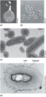
Capsules and Slime Layers
Capsules and slime layers are extracellular polysaccharide coats that protect cells and aid in attachment to surfaces. Capsules are tightly attached and visible with special stains, while slime layers are loosely attached and easily deformed.
Capsule: Dense, well-organized layer.
Slime Layer: Diffuse, easily deformed layer.
Functions: Protection from desiccation, immune evasion, and adherence to surfaces.
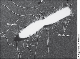
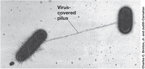
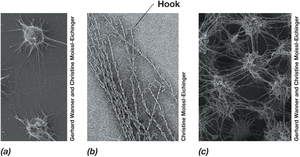
Cell Surface Structures: Fimbriae, Pili, and Hami
Microbial cells possess various surface structures that facilitate attachment, genetic exchange, and motility. Fimbriae are short, hair-like appendages, while pili are longer and involved in conjugation and electron transfer. Hami are unique to some archaea, enabling attachment to surfaces.
Fimbriae: Mediate attachment to surfaces and formation of biofilms.
Pili: Facilitate genetic exchange (conjugation) and can conduct electrons.
Hami: Specialized structures for surface attachment in archaea.
Endospores
Endospores are highly differentiated, dormant cells produced by certain Gram-positive bacteria. They are resistant to heat, radiation, chemicals, drying, and nutrient deprivation, enabling survival in harsh conditions.
Specialized Spores: Present in Bacillales and Clostridiales.
Survival Structures: Allow dispersal and persistence in unfavorable environments.
Flagella, Archaella, and Swimming Motility
Flagella (in bacteria) and archaella (in archaea) are motility structures that enable swimming through liquid environments. These appendages are anchored at one end and rotate to propel the cell.
Flagella: Long, thin appendages with various arrangements (polar, tufts).
Motility: Rotational speed is controlled by the proton motive force.
Chemotaxis
Chemotaxis is the directed movement of microorganisms in response to chemical stimuli. This behavior enhances access to nutrients and allows avoidance of harmful substances. Phototaxis is movement in response to light.
Taxis: General term for directed movement in response to stimuli.
Chemotaxis: Movement toward or away from chemicals.
Phototaxis: Movement in response to light.
