 Back
BackMicrobial Cell Structure and Function: Study Notes for Microbiology Students
Study Guide - Smart Notes
 Tailored notes based on your materials, expanded with key definitions, examples, and context.
Structure and Function of Bacterial Cells
Overview of the Microbial World
The microbial world encompasses a vast diversity of microscopic organisms, including Bacteria, Archaea, and Eukarya. Understanding their cell structure and function is fundamental to microbiology, as it reveals the unity and diversity of life and the mechanisms by which microbes interact with their environment.
Microbial Cell Structure and Function
The Cell Envelope
The cell envelope is a series of layered structures surrounding the cytoplasm, governing interactions with the environment. It is essential for maintaining cell integrity, shape, and protection against external stresses.
Cytoplasmic Membrane: Surrounds the cytoplasm and separates it from the environment. Its main function is selective permeability, allowing nutrients in and waste products out. Membrane proteins facilitate transport and energy metabolism.
Cell Wall: Provides rigidity and shape, preventing cell lysis due to osmotic pressure.
Outer Membrane: Present in gram-negative bacteria, adds an extra layer of protection.
S-layers: Protein or glycoprotein layers found in some Bacteria and Archaea.
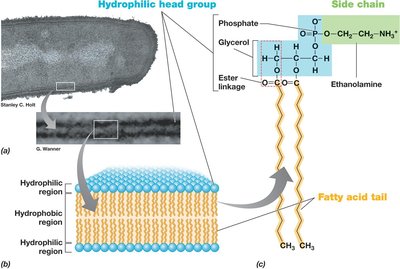
Cytoplasmic Membrane Structure
The cytoplasmic membrane is a phospholipid bilayer containing embedded proteins. It is 8–10 nm wide and consists of hydrophobic fatty acid tails and hydrophilic head groups (glycerol + phosphate + functional group).
Hydrophobic tails: Face inward, forming a barrier to water-soluble substances.
Hydrophilic heads: Face outward, interacting with the cytoplasm or external environment.
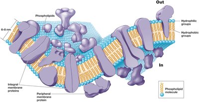
Membrane Proteins
Membrane proteins are crucial for transport and cellular processes:
Integral membrane proteins: Embedded within the membrane.
Transmembrane proteins: Span the entire membrane.
Peripheral membrane proteins: Loosely attached to the membrane surface.
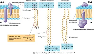
Archaeal Cytoplasmic Membranes
Archaea have unique membrane lipids with ether linkages (instead of ester) and isoprenoid chains. Major lipids include phosphoglycerol diethers and diphosphoglycerol tetraethers, which can form monolayers.
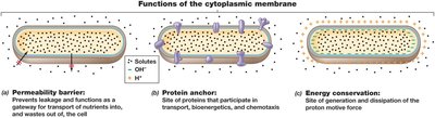
Functions of the Cytoplasmic Membrane
Permeability barrier: Prevents leakage and controls entry/exit of substances.
Protein anchor: Holds proteins involved in transport and energy metabolism.
Energy conservation: Site of generation and dissipation of the proton motive force.
Transporting Nutrients into the Cell
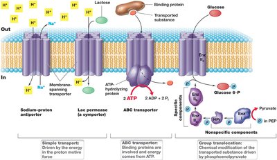
Microbial cells use active transport to accumulate solutes against concentration gradients. Three main mechanisms exist:
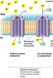
Simple transport: Uses transmembrane proteins, driven by proton motive force.
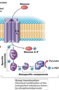
Group translocation: Involves a series of proteins; the transported substance is chemically modified (e.g., phosphotransferase system in E. coli).
ABC transporter system: Uses binding proteins, transmembrane transporters, and ATP-hydrolyzing proteins; over 200 systems exist for various substrates.
Simple Transport
Symport: Solute and H+ cotransported in one direction.
Antiport: Solute and H+ transported in opposite directions.
Group Translocation
Substance is chemically modified during transport.
Driven by energy-rich organic compounds (not proton motive force).
Example: Phosphotransferase system in E. coli (glucose, fructose, mannose).
ABC Transporter Systems
ATP-binding cassette transporters.
High substrate affinity; ATP drives uptake.
Ideal for nutrient-poor environments.
The Cell Wall
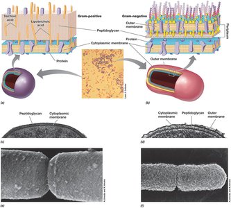
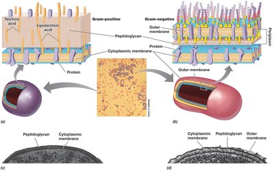
The cell wall is essential for withstanding osmotic pressure and maintaining cell shape. Most bacteria are classified as gram-positive or gram-negative based on cell wall structure and Gram stain reaction.
Gram-positive: Cytoplasmic membrane + thick cell wall.
Gram-negative: Cytoplasmic membrane, thin cell wall, outer membrane, periplasm.
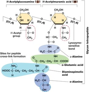
Peptidoglycan Structure
Peptidoglycan is a rigid polysaccharide layer providing strength. It consists of a sugar backbone (alternating N-acetylglucosamine and N-acetylmuramic acid) joined by β-1,4 linkages, with short peptides attached to N-acetylmuramic acid.
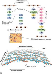
Peptidoglycan Arrangement
Strands run parallel around cell circumference.
Cross-linked by covalent peptide bonds.
Gram-negative: single layer; crosslinks between DAP and D-alanine.
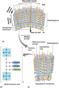
Gram-Positive Cell Envelope
Thick peptidoglycan wall (20–35 nm), up to 90% peptidoglycan.
Stabilized by peptide cross-links and interbridges (e.g., five glycines in Staphylococcus aureus).
Teichoic acids embedded in cell wall; lipoteichoic acids bound to membrane lipids.
Lysozyme cleaves glycosidic bonds; penicillin blocks peptide cross-links.
Archaeal Cell Walls
Lack peptidoglycan and typically lack outer membrane.
Most have S-layer (protein shell) instead of polysaccharide wall.
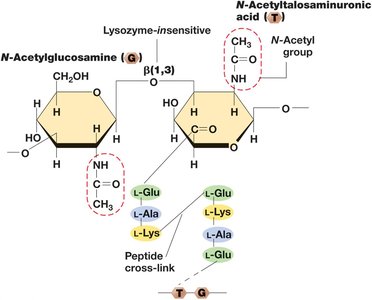
Methanogens have pseudomurein cell wall (alternating N-acetylglucosamine and N-acetyltalosaminuronic acid, β-1,3 bonds).
Resistant to lysozyme and penicillin.
LPS: The Outer Membrane
Gram-negative bacteria have an outer membrane composed of lipopolysaccharide (LPS), which is a second lipid bilayer external to the cell wall.
LPS: Contains core polysaccharide, O-polysaccharide, and lipid A (endotoxin).
Strengthened by ionic bonds to divalent cations (Ca2+, Mg2+).
Braun lipoprotein anchors outer membrane to peptidoglycan.
Porins facilitate solute transport.
Diversity of Cell Envelope Structure
S-Layers: Paracrystalline protein or glycoprotein structures, always outermost if present. Functions include strength, protection, shape, and adhesion.
Alternative Configurations: Some Bacteria and Archaea lack cell walls but have tough cytoplasmic membranes (e.g., Mycoplasmas, Thermoplasma).
Cell Surface Structures and Inclusions
Capsules and Slime Layers
Sticky polysaccharide coat outside cell envelope.
Capsule: tightly attached, visible with India ink.
Slime layer: loosely attached, easily deformed.
Functions: attachment, biofilm formation, infectivity, prevention of dehydration.
Fimbriae, Pili, and Hami
Pili: Thin protein filaments for surface attachment, biofilm formation, and genetic exchange (conjugation).
Fimbriae: Short pili mediating attachment.
Hami: Archaeal grappling hooks for biofilm formation.
Cell Inclusions
Energy reserves, carbon or phosphorus reservoirs, or special functions.
Enclosed by thin protein membrane, reducing osmotic stress.
Carbon storage polymers: PHB, PHA, glycogen.
Polyphosphate granules: Inorganic phosphate storage.
Elemental sulfur: Accumulates in periplasmic granules.
Carbonate minerals: Biomineralization of barium, strontium, magnesium.
Gas vesicles: Confer buoyancy.
Magnetosomes: Biomineralized magnetic iron oxides for magnetotaxis.
Endospores
Specialized, dormant cells resistant to heat, radiation, chemicals, drying, and nutrient deprivation.
Survival structures for dispersal and endurance.
Present only in some gram-positive bacteria (e.g., Bacillales, Clostridiales).
Formation triggered by nutrient limitation; germination occurs upon nutrient availability.
Structure: core, inner membrane, cortex, outer membrane, coat, exosporium; contains dipicolinic acid and SASPs.
Table: Differences Between Endospores and Vegetative Cells
Characteristic | Vegetative cell | Endospore |
|---|---|---|
Microscopic appearance | Nonrefractile | Refractile |
Calcium content | Low | High |
Dipicolinic acid | Absent | Present |
Enzymatic activity | High | Low |
Respiration rate | High | Low or absent |
Macromolecular synthesis | Present | Absent |
Heat resistance | Low | High |
Radiation resistance | Low | High |
Resistance to chemicals | Low | High |
Lysozyme | Sensitive | Resistant |
Water content | High, 80–90% | Low, 10–25% in core |
Small acid-soluble spore proteins | Absent | Present |
Cell Locomotion
Flagella, Archaella, and Swimming Motility
Flagella (Bacteria) and archaella (Archaea) are rotating appendages for swimming.
Arrangements: polar, tufts, lophotrichous, amphitrichous, peritrichous.
Structure: filament (flagellin), hook, basal body (motor).
Archaella are smaller, unrelated to flagella, and move by ATP hydrolysis.
Surface Motility
Twitching motility: Requires type IV pili, extends and retracts to pull cell forward.
Gliding motility: Smooth, continuous motion along cell axis, no external structures.
Chemotaxis and Other Forms of Taxis
Chemotaxis: Directed movement in response to chemicals.
Phototaxis: Response to light.
Aerotaxis: Response to oxygen.
Osmotaxis: Response to ionic strength.
Hydrotaxis: Response to water.
Eukaryotic Microbial Cells
The Nucleus and Cell Division
Eukaryotes contain a double membrane-enclosed nucleus and other organelles (mitochondria, Golgi complex, lysosomes, ER, microtubules, microfilaments).
Nucleus contains chromosomes; DNA is wound around histones forming nucleosomes.
Cell division: mitosis (two diploid cells), meiosis (four haploid gametes).
Mitochondria and Chloroplasts
Mitochondria: Site of respiration and ATP production; evolutionary roots in Bacteria.
Chloroplasts: Site of photosynthesis; contain chlorophyll and RuBisCO.
Endosymbiotic hypothesis: Organelles descended from bacterial cells associating with eukaryal hosts.
Other Eukaryotic Cell Structures
Cytoskeleton: Internal structural support (microtubules, microfilaments, intermediate filaments).
Endoplasmic Reticulum: Network of membranes; rough ER (ribosomes), smooth ER (lipid synthesis).
Golgi Complex: Modifies ER products.
Lysosomes: Contain digestive enzymes.
Flagella and Cilia: Motility organelles; structurally and functionally differ from prokaryotic flagella.
Additional info: These notes expand upon brief points in the original slides, providing definitions, examples, and academic context for each topic. All included images directly reinforce the explanations and are relevant to the adjacent paragraphs.
