 Back
BackMicrobial Cell Structure and Function: Study Notes for Microbiology Students
Study Guide - Smart Notes
 Tailored notes based on your materials, expanded with key definitions, examples, and context.
Structure and Function of Bacterial Cells
Overview of the Microbial World
The microbial world encompasses a vast diversity of organisms, including Bacteria, Archaea, and microbial Eukarya. Understanding their cell structure and function is fundamental to microbiology, as it reveals the unity and diversity of life and the mechanisms by which microbes interact with their environment.
Microbial Cell Structure and Function
The Cell Envelope
The cell envelope is a series of layered structures surrounding the cytoplasm, governing interactions with the environment. It includes the cytoplasmic membrane, cell wall, and, in some cases, an outer membrane or S-layer.
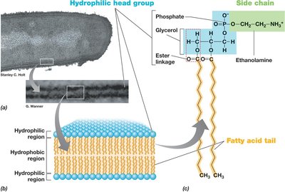
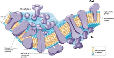
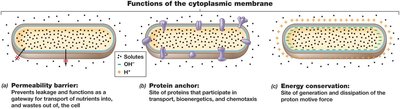
Cytoplasmic Membrane
The cytoplasmic membrane surrounds the cytoplasm and separates it from the environment. Its main function is selective permeability, allowing nutrients to enter and waste products to exit. Membrane proteins facilitate these reactions and play roles in energy metabolism.
Structure: Phospholipid bilayer containing embedded proteins, 8–10 nm wide. Hydrophobic fatty acid tails face inward, hydrophilic head groups face outward.
Membrane Proteins: Integral (embedded), transmembrane (span the membrane), and peripheral (loosely attached).
Functions: Permeability barrier, protein anchor, energy conservation (generation of proton motive force).
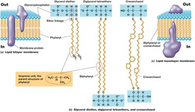
Archaeal Cytoplasmic Membranes
Ether linkages in phospholipids (vs. ester linkages in Bacteria/Eukarya).
Isoprenoid chains instead of fatty acids.
Lipid monolayers may form, providing stability in extreme environments.
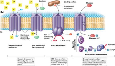
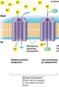
Transporting Nutrients into the Cell
Microbial cells use active transport to accumulate solutes against concentration gradients. Three main mechanisms exist:
Simple transport: Driven by proton motive force, involves a transmembrane protein.
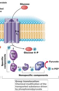
Group translocation: Substance is chemically modified during transport; energy from organic compounds (e.g., phosphotransferase system in E. coli).
ABC transporter systems: ATP-binding cassette; uses ATP and substrate-binding proteins for high-affinity uptake.

The Cell Wall
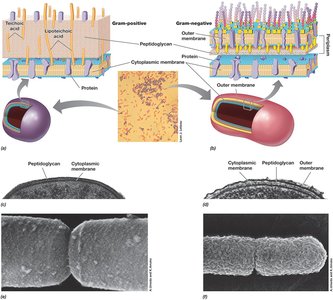
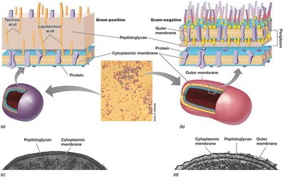
The cell wall provides structural strength and prevents cell lysis due to osmotic pressure. Most bacteria are classified as gram-positive or gram-negative based on cell wall structure and Gram stain reaction.
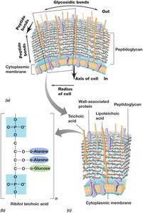
Gram-positive: Thick peptidoglycan cell wall, often with teichoic acids.
Gram-negative: Thin peptidoglycan layer, outer membrane, periplasmic space.
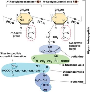
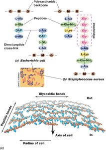
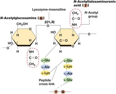
Peptidoglycan Structure
Rigid polysaccharide layer found in all Bacteria with a cell wall.
Glycan tetrapeptide: Alternating N-acetylglucosamine and N-acetylmuramic acid joined by β-1,4 linkages; short peptide attached to N-acetylmuramic acid.
Cross-linking: Covalent peptide bonds between glycan strands; varies between gram-positive and gram-negative bacteria.
Archaeal Cell Walls
Lack peptidoglycan; typically lack outer membrane.
S-layer: Protein shell providing structural support.
Pseudomurein: Found in some methanogens; similar to peptidoglycan but with β-1,3 glycosidic bonds and different sugars.
LPS: The Outer Membrane
Gram-negative bacteria possess an outer membrane composed of lipopolysaccharide (LPS), which is a major virulence factor and provides structural strength.
LPS: Contains core polysaccharide, O-polysaccharide, and lipid A (endotoxin).
Porins: Transmembrane proteins facilitating solute transport.
Periplasm: Space between cytoplasmic and outer membranes, housing extracellular proteins.
Diversity of Cell Envelope Structure
S-layers: Paracrystalline protein or glycoprotein structures, always outermost if present; provide strength, shape, and protection.
Alternative configurations: Some Bacteria and Archaea lack cell walls but have tough cytoplasmic membranes (e.g., Mycoplasmas, Thermoplasma).
Cell Surface Structures and Inclusions
Cell Surface Structures
Capsules and Slime Layers: Sticky polysaccharide coats outside the cell envelope; assist in attachment, biofilm formation, infectivity, and prevent dehydration.
Fimbriae and Pili: Protein filaments for attachment, biofilm formation, and genetic exchange (conjugation).
Hami: Archaeal grappling hooks for surface attachment and biofilm formation.
Cell Inclusions
Energy reserves: Poly-β-hydroxybutyric acid (PHB), poly-β-hydroxyalkanoate (PHA), glycogen.
Phosphorus and sulfur storage: Polyphosphate granules, elemental sulfur granules.
Carbonate minerals: Biomineralization of barium, strontium, magnesium.
Gas vesicles: Provide buoyancy.
Magnetosomes: Allow orientation within magnetic fields (magnetotaxis).
Endospores
Endospores are highly differentiated, dormant cells resistant to heat, radiation, chemicals, and desiccation. They are survival structures produced by some gram-positive bacteria (e.g., Bacillus, Clostridium).
Formation: Triggered by nutrient limitation; involves asymmetric cell division and forespore formation.
Structure: Multiple layers (core, inner membrane, cortex, coat, exosporium); contains dipicolinic acid and small acid-soluble spore proteins (SASPs).
Germination: Activation, germination, and outgrowth upon nutrient availability.
Characteristic | Vegetative cell | Endospore |
|---|---|---|
Microscopic appearance | Nonrefractile | Refractile |
Calcium content | Low | High |
Dipicolinic acid | Absent | Present |
Enzymatic activity | High | Low |
Respiration rate | High | Low or absent |
Macromolecular synthesis | Present | Absent |
Heat resistance | Low | High |
Radiation resistance | Low | High |
Resistance to chemicals | Low | High |
Lysozyme | Sensitive | Resistant |
Water content | High, 80–90% | Low, 10–25% in core |
Small acid-soluble spore proteins | Absent | Present |
Cell Locomotion
Flagella, Archaella, and Swimming Motility
Flagella: Long, thin appendages for swimming; arrangements include polar, lophotrichous, amphitrichous, peritrichous.
Structure: Filament (flagellin), hook, basal body (motor); rotates for movement.
Archaella: Smaller, unrelated to flagella, related to type IV pili; rotation driven by ATP hydrolysis.
Surface Motility
Twitching motility: Requires type IV pili; extension, attachment, retraction.
Gliding motility: Smooth, continuous motion along cell axis; involves intracellular protein tracks and motors.
Chemotaxis and Other Forms of Taxis
Chemotaxis: Directed movement in response to chemicals; "run and tumble" behavior in peritrichous bacteria.
Phototaxis: Response to light; photoreceptors interact with flagellar rotation proteins.
Aerotaxis: Response to oxygen; magnetotactic bacteria align with magnetic fields.
Osmotaxis and hydrotaxis: Response to ionic strength and water, respectively.
Eukaryotic Microbial Cells
The Nucleus and Cell Division
Nucleus: Double membrane-enclosed; contains chromosomes organized into nucleosomes (DNA + histones).
Nucleolus: Site of ribosomal RNA synthesis.
Cell division: Mitosis (two diploid cells), meiosis (four haploid gametes).
Mitochondria and Chloroplasts
Mitochondria: Site of respiration and ATP production; contains cristae and matrix.
Chloroplasts: Site of photosynthesis; contains thylakoids and stroma with RuBisCO.
Endosymbiotic hypothesis: Mitochondria and chloroplasts descended from bacterial cells, evidence includes circular DNA and bacterial-like ribosomes.
Other Eukaryotic Cell Structures
Cytoskeleton: Microtubules (tubulin), microfilaments (actin), intermediate filaments (keratin); maintain shape and facilitate motility.
Endoplasmic reticulum: Rough (ribosomes, protein synthesis), smooth (lipid synthesis, carbohydrate metabolism).
Golgi complex: Modifies ER products.
Lysosomes: Contain digestive enzymes.
Flagella and cilia: Motility organelles; structurally distinct from prokaryotic flagella.
Additional info: These notes expand on brief points with academic context, definitions, and examples to ensure completeness and clarity for microbiology students.
