 Back
BackMicrobial Cell Structure and Function: Study Notes for Microbiology Students
Study Guide - Smart Notes
 Tailored notes based on your materials, expanded with key definitions, examples, and context.
Structure and Function of Bacterial Cells
Introduction to the Microbial World
The microbial world encompasses a vast diversity of microscopic organisms, including bacteria, archaea, viruses, and microbial eukaryotes. Understanding their structure and function is fundamental to microbiology, as it reveals the unity and diversity of life and the mechanisms by which microbes interact with their environment.
Microbial Cell Structure and Function
The Cell Envelope
The cell envelope is a series of layered structures surrounding the cytoplasm, governing interactions with the environment and preserving cellular integrity. It consists of the cytoplasmic membrane, cell wall, and, in some cases, an outer membrane or S-layer.
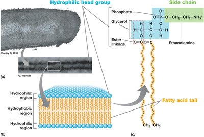
Cytoplasmic Membrane
Definition: The cytoplasmic membrane is a selectively permeable barrier that separates the cytoplasm from the external environment.
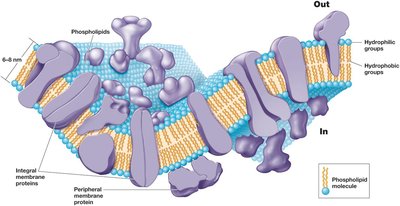
Structure: It is an 8–10 nm wide phospholipid bilayer containing embedded proteins. The bilayer has hydrophobic fatty acid tails facing inward and hydrophilic head groups exposed to the environment or cytoplasm.
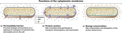
Function:
Selective permeability: Nutrients are transported in, and waste products are expelled.
Protein anchor: Holds proteins involved in transport and energy metabolism.
Energy conservation: Generates proton motive force for cellular energy.
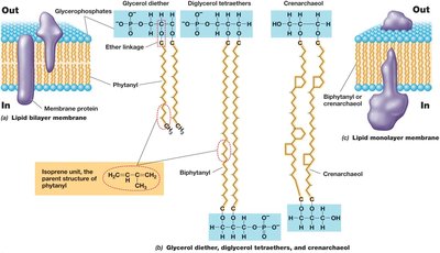
Archaeal Cytoplasmic Membranes
Differences: Archaeal membranes have ether linkages in phospholipids and isoprenoid chains, unlike the ester linkages and fatty acids in Bacteria and Eukarya.
Major lipids: Phosphoglycerol diethers and diphosphoglycerol tetraethers, which can form lipid monolayers.
Transporting Nutrients into the Cell
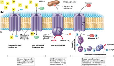
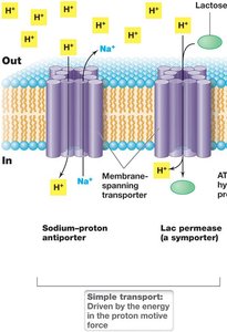
Microbial cells use active transport mechanisms to accumulate solutes against concentration gradients. Transporters are energy-driven and include three main types:
Simple transport: Uses a transmembrane protein and is driven by the proton motive force. Includes symport (solute and H+ cotransported) and antiport (solute and H+ transported in opposite directions).
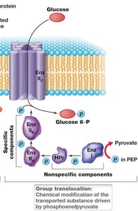
Group translocation: The transported substance is chemically modified during transport. The best-studied system is the phosphotransferase system in E. coli, which uses phosphoenolpyruvate as an energy source.
ABC transporter systems: Consist of a substrate-binding protein, transmembrane transporter, and ATP-hydrolyzing protein. These systems are ideal for organisms in nutrient-poor environments due to their high substrate affinity.

The Cell Wall
The cell wall provides structural strength and protects against osmotic pressure. Most bacteria are classified as gram-positive or gram-negative based on cell wall structure and Gram stain reaction.
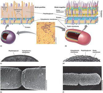
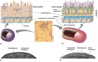
Gram-Positive vs. Gram-Negative Cell Envelopes
Gram-positive: Cytoplasmic membrane and thick peptidoglycan cell wall.
Gram-negative: Cytoplasmic membrane, thin peptidoglycan cell wall, outer membrane, and periplasmic space.
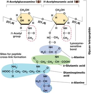
Peptidoglycan Structure
Definition: Peptidoglycan is a rigid polysaccharide layer found in all bacteria with a cell wall.
Composition: Alternating N-acetylglucosamine and N-acetylmuramic acid joined by β-1,4 linkages, with a short peptide attached to N-acetylmuramic acid.
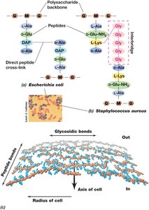
Cross-linking: Peptidoglycan strands are cross-linked by covalent peptide bonds, providing strength and rigidity.
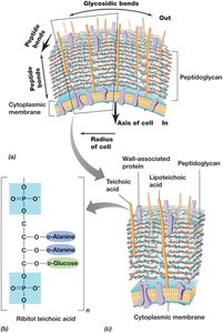
Gram-Positive Cell Envelope Features
Thick peptidoglycan wall, often stabilized by peptide interbridges.
Teichoic acids embedded in the cell wall, lipoteichoic acids covalently bound to membrane lipids.
Peptidoglycan can be destroyed by lysozyme; penicillin blocks peptide cross-link formation.
Archaeal Cell Walls
Lack peptidoglycan and typically lack an outer membrane.
Most have an S-layer (protein shell) instead of a polysaccharide wall.
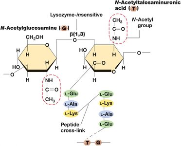
Methanogens have pseudomurein cell walls, which are similar to peptidoglycan but contain β-1,3 glycosidic bonds and L-stereoisomer amino acids.
LPS: The Outer Membrane
Gram-negative bacteria possess an outer membrane composed of a lipopolysaccharide (LPS) layer, which is a major virulence factor and adds structural strength.
LPS Structure: Contains a core polysaccharide, O-polysaccharide, and lipid A (endotoxin).
Function: Facilitates surface recognition, contains porins for solute transport, and anchors to peptidoglycan via Braun lipoprotein.
Diversity of Cell Envelope Structure
S-Layers: Paracrystalline protein or glycoprotein structures that provide strength, shape, and protection.
Alternative Configurations: Some bacteria and archaea lack cell walls but have tough cytoplasmic membranes (e.g., Mycoplasmas, Thermoplasma).
Cell Surface Structures and Inclusions
Cell Surface Structures
Capsules and Slime Layers: Sticky polysaccharide coats outside the cell envelope. Capsules are tightly attached; slime layers are loosely attached. Functions include attachment, biofilm formation, infectivity, and protection from desiccation.
Fimbriae and Pili: Protein structures that enable attachment to surfaces, biofilm formation, and genetic exchange (conjugation). Type IV pili support twitching motility.
Hami: Unique archaeal structures resembling grappling hooks, aiding in surface attachment and biofilm formation.
Cell Inclusions
Energy and Carbon Storage: Poly-β-hydroxybutyric acid (PHB), poly-β-hydroxyalkanoate (PHA), and glycogen are common storage polymers.
Polyphosphate, Sulfur, and Carbonate Minerals: Polyphosphate granules, elemental sulfur, and biomineralized carbonate minerals serve as reservoirs.
Gas Vesicles: Protein-bound structures that confer buoyancy to aquatic microbes.
Magnetosomes: Biomineralized magnetic iron oxides that allow magnetotaxis (migration along magnetic field lines).
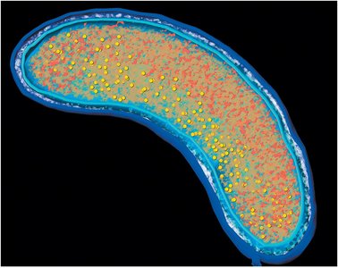
Endospores
Definition: Highly differentiated, dormant cells resistant to heat, radiation, chemicals, and desiccation.
Formation: Sporulation is triggered by nutrient limitation, resulting in a heat-resistant structure.
Structure: Multiple layers (core, inner membrane, cortex, outer membrane, coat, exosporium), contains dipicolinic acid and small acid-soluble spore proteins (SASPs).
Function: Survival and dispersal under unfavorable conditions; present only in some gram-positive bacteria.
Characteristic | Vegetative cell | Endospore |
|---|---|---|
Microscopic appearance | Nonrefractile | Refractile |
Calcium content | Low | High |
Dipicolinic acid | Absent | Present |
Enzymatic activity | High | Low |
Respiration rate | High | Low or absent |
Macromolecular synthesis | Present | Absent |
Heat resistance | Low | High |
Radiation resistance | Low | High |
Resistance to chemicals | Low | High |
Lysozyme | Sensitive | Resistant |
Water content | High, 80–90% | Low, 10–25% in core |
Small acid-soluble spore proteins | Absent | Present |
Cell Locomotion
Flagella, Archaella, and Swimming Motility
Flagella: Long, thin appendages anchored in the cell, functioning as rotating machines for swimming. Arrangements include polar, tufts, lophotrichous, amphitrichous, and peritrichous.
Structure: Composed of a filament (flagellin), hook, and basal body (motor).
Archaella: Smaller, unrelated to flagella, more closely related to type IV pili, and driven by ATP hydrolysis.
Surface Motility
Twitching Motility: Requires type IV pili, which extend, attach, and retract to pull the cell forward.
Gliding Motility: Smooth, continuous motion along the cell axis, involving intracellular protein tracks and adhesion proteins.
Chemotaxis and Other Forms of Taxis
Chemotaxis: Directed movement in response to chemical stimuli, involving "run and tumble" behavior in peritrichously flagellated bacteria.
Other Taxis: Osmotaxis (ionic strength), hydrotaxis (water), aerotaxis (oxygen), phototaxis (light), and magnetotaxis (magnetic fields).
Eukaryotic Microbial Cells
The Nucleus and Cell Division
Nucleus: Double membrane-enclosed organelle containing chromosomes. DNA is wound around histones, forming nucleosomes.
Cell Division: Mitosis produces two diploid daughter cells; meiosis produces four haploid gametes.
Mitochondria and Chloroplasts
Mitochondria: Site of respiration and ATP production in aerobic eukaryotes. Contains cristae (folded internal membranes) and matrix (innermost area).
Chloroplasts: Site of photosynthesis in phototrophic eukaryotes. Contains thylakoids (chlorophyll-containing discs) and stroma (with RuBisCO enzyme).
Endosymbiotic Origin: Mitochondria and chloroplasts descended from bacterial cells via symbiosis.
Other Eukaryotic Cell Structures
Cytoskeleton: Internal structural support composed of microtubules, microfilaments, and intermediate filaments.
Endoplasmic Reticulum and Golgi Complex: Networks for protein and lipid synthesis, modification, and transport.
Lysosomes: Membrane-enclosed compartments containing digestive enzymes.
Flagella and Cilia: Motility organelles structurally distinct from prokaryotic flagella, composed of microtubules and driven by ATP.
Example: The presence of mitochondria and chloroplasts in eukaryotic cells is evidence of endosymbiotic events, as these organelles contain their own DNA and ribosomes similar to bacteria.
Additional info: These notes expand on brief points by providing definitions, examples, and academic context to ensure completeness and clarity for exam preparation.
