 Back
BackMicrobial Cell Structure and Function – Study Notes
Study Guide - Smart Notes
 Tailored notes based on your materials, expanded with key definitions, examples, and context.
Overview of Microbial Cell Structures and Functions
This chapter introduces the fundamental structures and functions of microbial cells, focusing on the differences between prokaryotic and eukaryotic cells, and the specialized features that enable microorganisms to survive and thrive in diverse environments.
Cell Envelope/Membrane: Phospholipid bilayers that form the boundary of the cell.
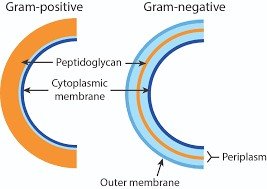
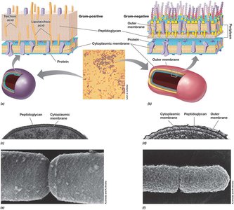
Cell Walls: Composed of peptidoglycan; thick in Gram-positive (G+), thin in Gram-negative (G-).
Capsules: Polysaccharide layers that act as virulence factors by preventing phagocytosis.
Fimbriae: Structures for attachment to surfaces.
Pili: Involved in conjugation (genetic exchange).
Flagella: Enable motility.
Inclusions: Storage of nutrients and energy.
Endospores: Survival structures under harsh conditions.
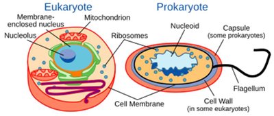
The Cell Envelope
Structure and Components
The cell envelope is a series of layered structures that surround the cytoplasm and mediate interactions with the environment. It is essential for maintaining cell integrity and mediating transport.
Cytoplasmic membrane: Present in all cells; primary permeability barrier.
Cell wall: Present in most bacteria; provides rigidity and shape.
Outer membrane: Found only in Gram-negative bacteria.
S-layers: Protein or glycoprotein layers found in some bacteria and archaea.
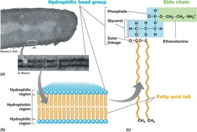
Cell (Cytoplasmic) Membrane Structure
Phospholipid Bilayer
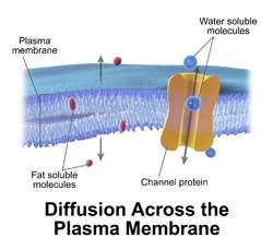
The cytoplasmic membrane is a phospholipid bilayer containing embedded proteins. It is 8–10 nm wide and separates the cytoplasm from the environment, maintaining selective permeability.
Hydrophobic region: Fatty acid tails face inward, forming a hydrophobic core.
Hydrophilic region: Glycerol, phosphate, and functional groups face outward.
Membrane proteins: Integral (embedded), transmembrane (span the membrane), and peripheral (loosely attached).
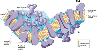
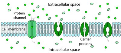
Selective Permeability
The cytoplasmic membrane is selectively permeable, allowing nutrients to enter and waste products to exit. Membrane proteins facilitate transport and energy metabolism.
Passive transport: Diffusion and facilitated diffusion via channel and carrier proteins.
Active transport: Requires energy to move substances against their concentration gradient.
Variations Across Domains
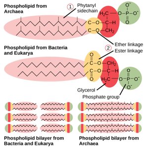
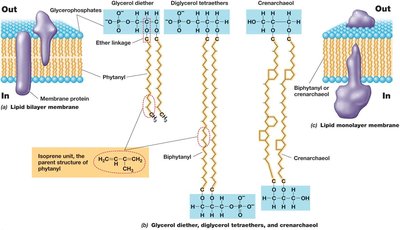
Membrane lipid composition differs among the three domains of life, affecting chemical stability and adaptation to extreme environments.
Bacteria & Eukarya: Fatty acid chains linked to glycerol via ester bonds.
Archaea: Isoprene chains linked via ether bonds, forming monolayers or bilayers; more chemically stable.
The Cell Wall
Function and Composition
The cell wall provides structural support, maintains cell shape, and prevents lysis due to osmotic pressure. Most bacteria have cell walls composed of peptidoglycan, while archaea have distinct wall structures.
Gram-positive: Thick peptidoglycan layer, teichoic acids.
Gram-negative: Thin peptidoglycan, outer membrane, periplasmic space.
Acid-fast bacteria: Waxy cell walls with mycolic acid (e.g., Mycobacterium).
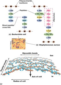
Peptidoglycan Structure
Peptidoglycan is a rigid polysaccharide layer unique to bacteria, consisting of glycan chains cross-linked by peptides.
Sugar backbone: Alternating N-acetylglucosamine (NAG) and N-acetylmuramic acid (NAM) joined by β-1,4 linkages.
Peptide side chains: Short peptides attached to NAM, varying among species.
Cross-linking: Covalent peptide bonds provide strength; Gram-positive bacteria often have peptide interbridges.


Gram-Positive vs. Gram-Negative Cell Walls
Gram-positive: Up to 90% peptidoglycan, multiple layers, teichoic acids for stability.
Gram-negative: Thin peptidoglycan, outer membrane with lipopolysaccharide (LPS), periplasmic space.
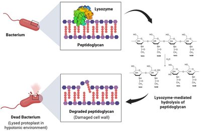
Destruction of Peptidoglycan
Lysozyme: Enzyme that cleaves glycosidic bonds in peptidoglycan, found in human secretions.
Penicillin: Antibiotic that blocks peptide cross-link formation, weakening the cell wall.
Archaeal Cell Walls
No peptidoglycan: Instead, S-layers (protein shells) or pseudomurein in some methanogens.
Pseudomurein: Alternating N-acetylglucosamine and N-acetyltalosaminuronic acid, β-1,3 linkages, resistant to lysozyme and penicillin.
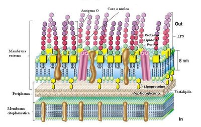
Gram-Negative Outer Membrane and LPS
Lipopolysaccharide (LPS) Layer
The outer membrane of Gram-negative bacteria contains LPS, which is important for surface recognition, virulence, and structural integrity.
Lipid A: Endotoxin component, toxic to animals.
Core polysaccharide and O-polysaccharide: Contribute to antigenic specificity.
Porins: Protein channels for solute transport.
Periplasm: Space between cytoplasmic and outer membranes, containing various proteins.
Diversity of Cell Envelope Structure
S-Layers
S-layers are paracrystalline protein or glycoprotein layers found in some Bacteria and Archaea, providing strength, protection, and mediating cell interactions.
Wall-less Microbes
Mycoplasma (Bacteria): Lack cell walls, have sterol-rich membranes.
Thermoplasma (Archaea): Lack cell walls, have tough membranes.
Cell Surface Structures – Virulence Factors
Capsules and Slime Layers
Capsules and slime layers are polysaccharide coats outside the cell envelope, contributing to virulence by aiding attachment, biofilm formation, and protection from desiccation and host defenses.
Capsule: Tightly attached, visible with special stains.
Slime layer: Loosely attached, easily deformed.
Pili and Fimbriae
Pili: Protein filaments for attachment, biofilm formation, and conjugation (genetic exchange).
Fimbriae: Shorter, used mainly for attachment to surfaces.
Type IV pili: Enable twitching motility.
Hamus/Hami
Specialized grappling hook-like structures in some Archaea for attachment and biofilm formation.
Cell Inclusions
Storage and Special Functions
Inclusions are intracellular storage bodies for energy reserves, carbon, phosphorus, and other minerals, reducing osmotic stress.
Carbon storage: Poly-β-hydroxybutyric acid (PHB), glycogen.
Polyphosphate granules: Inorganic phosphate storage.
Sulfur granules: Elemental sulfur storage.
Carbonate minerals: Biomineralization of barium, strontium, magnesium.
Gas vesicles: Provide buoyancy in aquatic bacteria (e.g., cyanobacteria).
Magnetosomes: Allow orientation within magnetic fields (magnetotaxis).
Endospores
Structure and Function
Endospores are highly resistant, dormant structures formed by some Gram-positive bacteria (e.g., Bacillus, Clostridium) to survive extreme conditions.
Formation (sporulation): Triggered by nutrient limitation; involves differentiation from vegetative cell to spore.
Germination: Activation, germination, and outgrowth upon nutrient availability.
Structure: Contains dipicolinic acid, Ca2+, and small acid-soluble spore proteins (SASPs) for DNA protection.
Bacterial Motility
Types of Motility
Swimming: Movement in liquid via flagella.
Twitching: Surface movement using type IV pili.
Gliding/Sliding: Surface movement without external appendages.
Swarming: Coordinated group movement, often increasing resistance to antimicrobials.
Flagella and Archaella
Flagella (Bacteria) and archaella (Archaea) are rotary appendages for swimming. Arrangements include polar, lophotrichous, amphitrichous, and peritrichous. The flagellum consists of a filament, hook, and basal body.
Taxis – Directed Movement
Chemotaxis
Bacteria and Archaea move toward or away from chemical stimuli using a 'run and tumble' mechanism (peritrichous) or reversal (polar). Chemoreceptors detect changes in concentration over time.
Other Forms of Taxis
Phototaxis: Movement toward light.
Aerotaxis: Movement toward or away from oxygen.
Magnetotaxis: Orientation and movement along magnetic fields.
Osmotaxis/Hydrotaxis: Response to ionic strength or water.
Endosymbiotic Origin of Organelles
The endosymbiotic hypothesis proposes that mitochondria and chloroplasts originated from free-living bacteria that established symbiotic relationships with ancestral eukaryotic cells. Evidence includes the presence of circular DNA and bacterial-like ribosomes in these organelles.
