 Back
BackMicrobial Cell Structure: Cell Membranes and Cell Walls in Prokaryotes
Study Guide - Smart Notes
 Tailored notes based on your materials, expanded with key definitions, examples, and context.
Cell Envelope Structure in Prokaryotes
Definition and Components
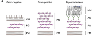
The cell envelope is a series of layered structures that surround the cytoplasm of prokaryotic cells, governing interactions with the environment. It typically consists of the cytoplasmic membrane, cell wall, and, in some cases, an outer membrane or S-layer.
Cytoplasmic membrane: Innermost boundary, responsible for selective permeability.
Cell wall: Provides structural support and shape.
Outer membrane: Present in Gram-negative bacteria, adds an extra layer of protection.
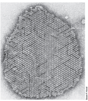
S-layer: Paracrystalline protein or glycoprotein shell, outermost if present.
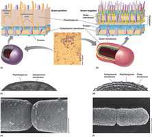
Gram-positive and Gram-negative bacteria have distinct cell envelope architectures.

Cytoplasmic Membrane
Structure and Function
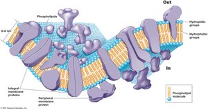
The cytoplasmic membrane surrounds the cytoplasm and separates it from the environment. Its main function is selective permeability, allowing nutrients to enter and waste products to exit. Membrane proteins facilitate transport and energy metabolism.
Phospholipid bilayer: 8–10 nm wide, contains embedded proteins.
Hydrophobic tails: Fatty acids oriented inward.
Hydrophilic heads: Glycerol, phosphate, and functional groups exposed to the environment or cytoplasm.
Membrane Strengthening Agents
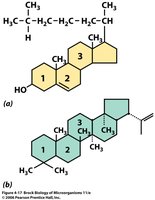
Some membranes contain strengthening agents:
Sterols: Found in eukaryotes.
Hopanoids: Found in bacteria.
Archaeal Cytoplasmic Membranes
Archaea have unique membrane structures:
Can be lipid bilayers or monolayers.
Contain isoprenoid chains (e.g., phytanyl) instead of fatty acids.
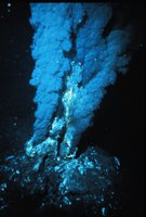
Adapted for extreme environments, such as high temperature.

Proteins in the Cell Membrane
Types and Functions
Membrane proteins are essential for:
Transport: Moving substances across the membrane.
Energy conservation: Involved in metabolic processes.
Regulation: Controlling cellular activities.
Motility: Facilitating movement.
Cell Walls of Prokaryotes
Structure and Function
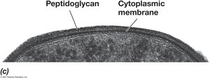
The cell wall provides mechanical strength, shape, and protection. It is primarily composed of peptidoglycan in bacteria.
Gram-positive: Thick peptidoglycan layer.
Gram-negative: Thin peptidoglycan layer, outer membrane present.

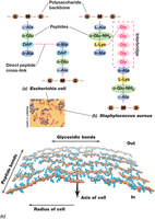
Peptidoglycan Structure
Peptidoglycan is a polymer of sugars and amino acids forming a mesh-like layer outside the cytoplasmic membrane.
Monomers: N-acetylglucosamine (NAG) and N-acetylmuramic acid (NAM).
Glycosidic bonds between NAG-NAM (lysozyme sensitive).
Peptide bonds between NAM-NAM (protease insensitive).
Repeating units:
Cross-linking varies: DAP-Ala (Gram-negative), Lys-Gly (Gram-positive).

Gram-Positive Cell Wall
Gram-positive bacteria have a thick peptidoglycan layer (25–35 nm), with embedded molecules such as teichoic acids and cell wall proteins.
High mechanical strength.
Protective function.
Protein anchor.
Gram-Negative Cell Wall
Gram-negative bacteria possess a thin peptidoglycan layer and an outer membrane containing lipopolysaccharides (LPS), which are important for surface recognition and virulence.
Outer membrane is asymmetric: LPS (outer), lipids (inner).
Contains porins for transport.
Periplasmic space between membranes (~15 nm wide).
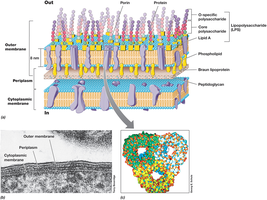
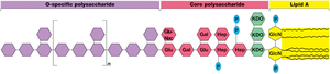
Lipopolysaccharides (LPS)
LPS consists of a core polysaccharide, O-polysaccharide, and lipid A (endotoxin). LPS replaces most phospholipids in the outer half of the outer membrane and is anchored by Braun lipoprotein.
Ionic bonds to divalent cations (Ca2+, Mg2+) add strength.
Lipid A is the toxic component (endotoxin).
Archaeal Cell Walls
Some Archaea possess pseudomurein, a chemical analog to peptidoglycan. Pseudomurein contains NAT instead of NAM, is lysozyme insensitive, and has peptide cross-links with only L-amino acids.
Variations on Envelope Structure
S-layer: Paracrystalline protein or glycoprotein shell, outermost layer if present.
Mycobacterial cell wall: Mycolic acid outer membrane (waxy), found only in Gram-positive Mycobacteria.
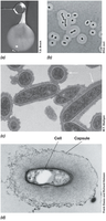
Capsules and Slime Layers
Functions and Structure
Capsules and slime layers are extracellular layers composed of polysaccharides and some proteins. They assist in attachment, biofilm formation, infectivity, and protection against desiccation and immune detection.
Fimbriae and Pili
Structure and Function
Pili are thin, filamentous protein structures (2–10 nm wide) that enable bacteria to adhere to surfaces, form biofilms, and facilitate genetic exchange (conjugation). Fimbriae are shorter pili that mediate attachment.
Produced by all Gram-negatives and many Gram-positives.
Electrically conductive pili can conduct electrons.
Cell Inclusions
Gas Vesicles
Gas vesicles are conical-shaped, gas-filled structures made of two proteins, conferring buoyancy to cells such as cyanobacteria.
Endospores
Structure and Function
Endospores are highly differentiated, dormant cells resistant to heat, radiation, chemicals, and desiccation. They serve as survival structures for dispersal and are present only in some Gram-positive bacteria (e.g., Bacillales and Clostridiales).
Formation triggered by nutrient limitation (sporulation).
Can remain dormant for years; germination occurs rapidly when nutrients are available.
Three steps: activation, germination, outgrowth.
Structure: core, inner membrane, cortex, outer membrane, endospore coat, exosporium.
Contains dipicolinic acid and small acid-soluble spore proteins (SASPs) for DNA protection and energy source.
Sporulation involves asymmetric cell division and forespore formation, with over 200 sporulation-specific genes in Bacillus subtilis.
Feature | Endospore | Vegetative Cell |
|---|---|---|
Resistance | High (heat, chemicals, radiation) | Low |
Metabolic Activity | Dormant | Active |
Structure | Multiple layers, dipicolinic acid, SASPs | Single membrane, no special layers |
Summary Table: Comparison of Cell Wall Structures
Type | Main Components | Special Features |
|---|---|---|
Gram-positive | Thick peptidoglycan, teichoic acids | High strength, protein anchor |
Gram-negative | Thin peptidoglycan, outer membrane (LPS) | Periplasmic space, porins, endotoxin |
Archaea | Pseudomurein or S-layer | Lysozyme insensitive, L-amino acids |
Mycobacteria | Mycolic acid, peptidoglycan | Waxy outer membrane, acid-fast |
