 Back
BackMicrobial Cell Structure: Cytoplasmic Membranes and Cell Walls
Study Guide - Smart Notes
 Tailored notes based on your materials, expanded with key definitions, examples, and context.
Cytoplasmic Membrane Structure and Function
Overview of the Cytoplasmic Membrane
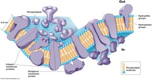
The cytoplasmic membrane is a highly selective permeability barrier that separates the interior of the cell from its external environment. It is composed primarily of lipids and proteins, forming a dynamic and fluid structure essential for cellular function.
Fluid Mosaic Model: The membrane is described as a 'fluid mosaic' due to the lateral movement of lipids and proteins within the bilayer.
Selective Permeability: Only certain molecules can pass through freely, while others require specific transport mechanisms.
Phospholipid Bilayer in Bacteria and Eukarya
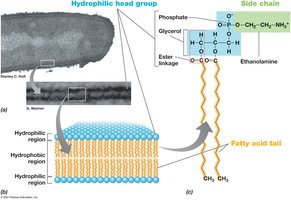
In Bacteria and Eukarya, the cytoplasmic membrane consists of a phospholipid bilayer with ester linkages connecting fatty acids to glycerol. This structure provides both fluidity and stability to the membrane.
Hydrophilic Head Groups: Face outward, interacting with aqueous environments.
Hydrophobic Fatty Acid Tails: Face inward, forming a barrier to most water-soluble substances.
Archaeal Membranes
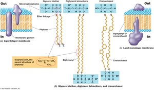
Archaea possess unique membrane lipids. Instead of fatty acids, they contain phytanyl chains attached to glycerol via ether linkages. Some archaeal membranes form a monolayer using biphytanyl, which increases resistance to extreme conditions.
Ether Linkages: More chemically stable than ester linkages, contributing to the resilience of Archaea in harsh environments.
Monolayer Membranes: Found in some Archaea, providing additional stability against heat and denaturation.
Other Membrane Lipids
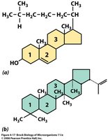
Additional lipids in the cytoplasmic membrane, such as sterols (e.g., cholesterol in Eukarya) and hopanoids (in Bacteria), help strengthen and stabilize the membrane.
Sterols: Found mainly in eukaryotic membranes, they modulate fluidity and permeability.
Hopanoids: Bacterial analogs of sterols, contributing to membrane rigidity.
Functions of the Cytoplasmic Membrane
The cytoplasmic membrane performs several critical functions necessary for cell survival and activity:
Permeability Barrier: Prevents leakage and acts as a gateway for nutrient and waste transport.
Protein Anchor: Provides a site for proteins involved in transport, bioenergetics, and chemotaxis.
Energy Conservation: Site of generation and dissipation of the proton motive force, essential for ATP synthesis.

Transport Across the Cytoplasmic Membrane
Types of Active Transport Events
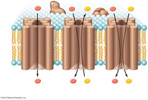
Transport across the membrane can occur via several mechanisms, including uniporters, symporters, and antiporters:
Uniporter: Transports a single type of molecule in one direction.
Symporter: Moves two substances in the same direction simultaneously.
Antiporter: Exchanges one substance for another across the membrane.
The Three Classes of Transport Systems
Microbial cells utilize three main classes of transport systems to move substances across the cytoplasmic membrane:
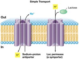
Simple Transport: Driven by the energy of the proton motive force.
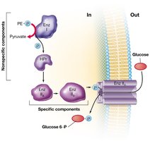
Group Translocation: Chemical modification of the transported substance driven by phosphoenolpyruvate (PEP).
ABC Transporters (ATP-Binding Cassette): Utilize ATP hydrolysis to drive transport.
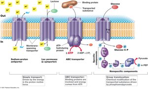
Examples of Simple Transport
Simple transport systems include antiporters and symporters, such as the sodium-proton antiporter and the lactose-proton symporter (Lac permease).
Sodium-Proton Antiporter: Exchanges Na+ for H+ across the membrane.
Lac Permease: Transports lactose into the cell along with H+.
Example of Group Translocation
Group translocation involves the chemical modification of a molecule as it is transported into the cell. The classic example is the phosphotransferase system (PTS) for glucose uptake in bacteria.
Phosphotransferase System (PTS): Glucose is phosphorylated during transport, using energy from phosphoenolpyruvate (PEP).
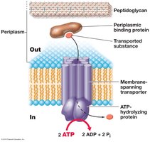
Mechanism of an ABC Transporter
ABC transporters use the energy from ATP hydrolysis to transport substances across the membrane. They are characterized by the presence of a periplasmic binding protein, a membrane-spanning transporter, and an ATP-hydrolyzing protein.
Binding Protein: Captures the substrate and delivers it to the transporter.
ATP Hydrolysis: Provides the energy for transport.
Generation and Use of Proton Motive Force
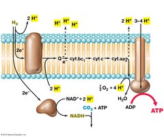
The electron transport chain in the cytoplasmic membrane generates a proton motive force (PMF), which is used by ATP synthase to produce ATP. This process is central to cellular energy metabolism.
Electron Transport Chain: Transfers electrons and pumps protons across the membrane, creating an electrochemical gradient.
ATP Synthase: Utilizes the PMF to synthesize ATP from ADP and inorganic phosphate.
Cell Wall Structure and Function
General Structure and Function
The cell wall is a rigid structure located outside the cytoplasmic membrane. It provides protection, maintains cell shape, prevents osmotic lysis, and mediates interactions with the environment. Its composition varies among Bacteria, Archaea, and Eukarya.
Major Components: Proteins, glycoproteins, and polysaccharides.
Special Cases: Some cells, such as Mycoplasma and Chlamydia, lack a cell wall.
Eukaryotic Cell Walls
Eukaryotic microorganisms exhibit diverse cell wall compositions:
Diatoms: Cell walls composed of silica (SiO2), protein, and polysaccharide.
Algae: Cellulose is the primary component.
Fungi: Chitin and glycoproteins are predominant.
Bacterial Cell Walls
Bacterial cell walls are primarily composed of peptidoglycan, except in Mycoplasma and Chlamydia (which lack cell walls) and Planctomycetes (which have protein cell walls). Peptidoglycan is a unique polymer that provides structural integrity.
Peptidoglycan: Found in nearly all Bacteria, distinguishing them from Archaea and Eukarya.
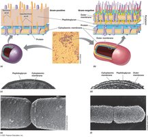
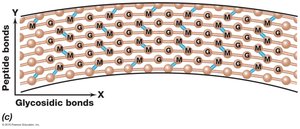
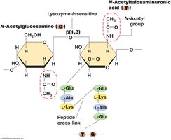
Peptidoglycan Structure and Gram Staining
Peptidoglycan is composed of repeating units of N-acetylglucosamine (NAG) and N-acetylmuramic acid (NAM) linked by β-1,4-glycosidic bonds, with peptide crosslinks providing additional strength. The thickness of the peptidoglycan layer distinguishes Gram-positive from Gram-negative bacteria.
Gram-Positive: Thick peptidoglycan layer (up to 90% of cell wall).
Gram-Negative: Thin peptidoglycan layer (about 10% of cell wall), with an additional outer membrane.
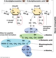
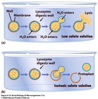
Lysozyme and Peptidoglycan Degradation
Lysozyme is an enzyme found in animal secretions that destroys peptidoglycan by cleaving β-1,4-glycosidic bonds between NAG and NAM. It serves as a major defense mechanism against bacterial infection.
Effect: Disrupts the cell wall, leading to cell lysis in hypotonic environments.
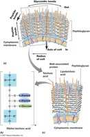
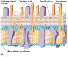
Gram-Positive Cell Envelope
The Gram-positive cell envelope consists of a thick peptidoglycan layer and cytoplasmic membrane. Teichoic acids and lipoteichoic acids are important components, contributing to the cell wall's negative charge and binding of cations.
Teichoic Acids: Polymers containing glycerol phosphate or ribitol phosphate residues.
Lipoteichoic Acids: Teichoic acids covalently linked to membrane lipids.
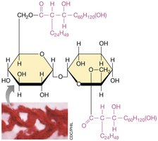
Mycolic Acids in Mycobacterium
Some Gram-positive bacteria, such as Mycobacterium, contain mycolic acids in their cell walls. These hydrophobic molecules contribute to the formation of the cord factor and provide resistance to desiccation and chemical damage.
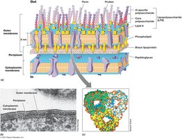
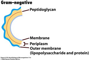
Gram-Negative Cell Envelope
The Gram-negative cell envelope is more complex, consisting of a thin peptidoglycan layer, an outer membrane, and a periplasmic space. The outer membrane contains lipopolysaccharide (LPS), which is important for structural integrity and pathogenicity.
LPS Components: Lipid A (toxic), core polysaccharide, and O-specific polysaccharide.
Porins: Proteins that allow the passage of small molecules through the outer membrane.
Archaeal Cell Walls
Archaeal cell walls are diverse and may contain pseudomurein, protein, glycoprotein, or polysaccharide. Pseudomurein is similar to peptidoglycan but contains different sugars and β-1,3-glycosidic bonds, making it insensitive to lysozyme.
Pseudomurein: Found in some methanogenic Archaea, such as Methanobacterium.
Polysaccharide Walls: Found in other Archaea, such as Halococcus.
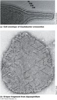
S-Layers
The S-layer is a two-dimensional array of protein or glycoprotein that forms a selective sieve on the surface of some Archaea and Bacteria. It provides structural support and protection against environmental stress.
Viral Envelope
Some viruses possess an envelope derived from the host cell membrane. This lipid bilayer surrounds the viral capsid and plays a role in viral entry and immune evasion.
Origin: Acquired from the host during viral budding.
Function: Facilitates attachment and fusion with host cells.
