 Back
BackMicrobial Cell Structure, Division, Biofilm Formation, and Antibiotic Targets
Study Guide - Smart Notes
 Tailored notes based on your materials, expanded with key definitions, examples, and context.
Bacterial Cell Division and the Divisome
Divisome Complex and Its Role
The divisome is a protein complex essential for bacterial cell division, ensuring the formation of two identical daughter cells. It forms late in the cell cycle, after DNA replication, and orchestrates the constriction of the cell membrane and synthesis of new peptidoglycan cell wall at the division site.
FtsZ: Forms the Z-ring at the cell center, uses GTP for energy, and initiates septum formation.
Min Proteins (MinC, MinD, MinE): Prevent division at incorrect locations, guiding FtsZ to the cell midpoint.
FtsK: Assists in chromosome separation during division.
The divisome ensures equal partitioning of cellular contents and chromosomes, resulting in two identical daughter cells.
Cytoskeleton and Cell Shape
Bacterial Cytoskeleton Proteins
Bacteria possess cytoskeletal proteins analogous to those in eukaryotes, which are crucial for maintaining cell shape and structure.
MreB: Similar to actin, forms filaments under the membrane, recruits cell wall synthesis proteins, and determines rod shape. Inactivation leads to cocci shape.
CrvA: In Vibrio species, adds peptidoglycan to one side, causing cell curvature.
Crescentin: Similar to keratin, forms filaments on the concave side, creating crescent-shaped cells.
These proteins provide evolutionary evidence for relatedness between domains of life.
Peptidoglycan Biosynthesis and Cell Wall Growth
Mechanisms of Peptidoglycan Synthesis
Peptidoglycan synthesis is vital during cell division and elongation, involving two main complexes:
Divisome: Produces peptidoglycan during division.
Elongasome: Produces peptidoglycan during cell growth.
Rod-shaped bacteria grow their wall along the cell length, while cocci expand outward from the FtsZ ring.
Insertion of New Peptidoglycan
Break old cell wall: Autolysins degrade existing peptidoglycan, creating space for new material.
Transport new building blocks: Bactoprenol carries peptidoglycan precursors (Lipid I, Lipid II) across the membrane. Flippase flips bactoprenol. Bacitracin blocks bactoprenol.
Insert new wall: Transglycosylase adds sugar chains, forming glycosidic bonds. Transpeptidation cross-links peptidoglycan, strengthening the wall. Penicillin inhibits transpeptidation.
Endospore Formation and Germination
Endospore Formation
Some Gram-positive bacteria, such as Bacillus and Clostridium, form endospores under stress for survival.
Environmental stress activates Spo0A.
Spo0A triggers over 500 genes.
Asymmetric division produces a small endospore and a larger mother cell.
Endospore develops a thick coat, dipicolinic acid, and extreme resistance.
Endospore Germination
Endospores are dormant and metabolically inactive, surviving for years. Upon favorable conditions, they undergo:
Activation
Germination
Outgrowth
Result: Return to vegetative cell.
Biofilm Formation and Regulation
Stages of Biofilm Formation
A biofilm is a community of bacteria attached to a surface, such as dental plaque or medical devices. Formation occurs in four stages:
Attachment
Colonization
Development
Dispersal
Attachment Mechanisms
Initial attachment via random collisions, flagella, pili, and cell surface proteins.
Cells lose flagella and become non-motile after attachment.
Regulation by c-di-GMP
Biofilm formation is regulated by cyclic di-GMP (c-di-GMP), a signaling molecule that controls the switch between motile and biofilm states.
Low c-di-GMP | High c-di-GMP |
|---|---|
Motile cells | Biofilm formation |
Reduces flagella activity
Promotes extracellular matrix production
Biofilm Examples
Pseudomonas aeruginosa: Forms strong biofilms at high cell density, high antibiotic resistance, releases DNA into matrix.
Vibrio cholerae: Biofilm formation activated at low cell density, repressed at high density via quorum sensing and HapR repressor.
Antibiotic Targets and Resistance
Major Antibiotic Targets
Cell membrane
Cell wall
DNA replication
RNA synthesis
Ribosomes
DNA gyrase
Examples of Antibiotic Action
DNA Replication: Quinolones inhibit DNA gyrase.
Protein Synthesis: Bacterial ribosome (70S) targeted by puromycin and aminoglycosides; eukaryotic ribosome (80S) is not affected.
Cell Membrane: Daptomycin forms pores; polymyxins target LPS layer.
Cell Wall: β-lactam antibiotics (penicillin, cephalosporins) block transpeptidation; vancomycin blocks precursor; bacitracin blocks bactoprenol.
Antibiotic Resistance Mechanisms
Drug target modification
Drug inactivation (e.g., β-lactamase)
Efflux pumps (e.g., AcrAB-TolC in E. coli)
Metabolic bypass (e.g., MRSA with mecA gene)
Persistence, Dormancy, and Toxin–Antitoxin Systems
Persistence and Dormancy
Some bacteria survive antibiotics by entering a dormant state (persister cells), which are genetically identical but temporarily inactive. After treatment, they can resume growth and cause recurring infections.
Examples: Mycobacterium tuberculosis, Pseudomonas aeruginosa
Toxin–Antitoxin Modules
Bacteria possess genes regulating dormancy through toxin-antitoxin systems:
Toxin: Stops cell growth
Antitoxin: Neutralizes toxin
Example: HipA–HipB system
Stringent Response
Response to Amino Acid Starvation
During amino acid starvation, bacteria activate the stringent response:
Ribosome stalls
RelA produces alarmone (p)ppGpp
Stringent response activates
Decreases rRNA and tRNA synthesis
Reduces protein production
Slows DNA replication
Stops cell division
Result: Cell enters dormancy and resumes growth once conditions improve.
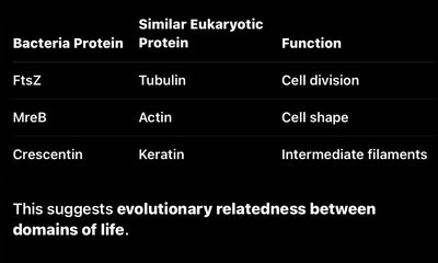
Summary Table: Bacterial Cytoskeleton Proteins and Functions
Bacteria Protein | Similar Eukaryotic Protein | Function |
|---|---|---|
FtsZ | Tubulin | Cell division |
MreB | Actin | Cell shape |
Crescentin | Keratin | Intermediate filaments |
This suggests evolutionary relatedness between domains of life.
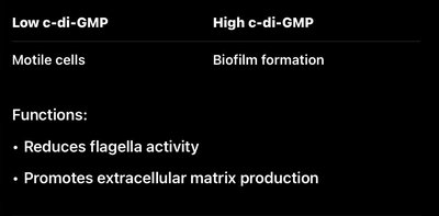
Summary Table: c-di-GMP Regulation of Biofilm Formation
Low c-di-GMP | High c-di-GMP |
|---|---|
Motile cells | Biofilm formation |
Reduces flagella activity
Promotes extracellular matrix production
Key Equations
Transpeptidation Reaction (Peptidoglycan Cross-linking)
The transpeptidation reaction forms peptide cross-links between peptidoglycan chains:
Stringent Response Alarmone Synthesis
RelA synthesizes (p)ppGpp from GTP and ATP:
Additional info:
Tables and images were recreated and expanded for clarity and completeness.
Academic context was added to ensure self-contained study notes.
