 Back
BackLecture 12
Study Guide - Smart Notes
 Tailored notes based on your materials, expanded with key definitions, examples, and context.
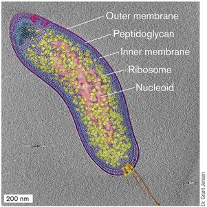
Microbial Cell Structure
Overview of Microbial Cell Components
Microbial cells possess a variety of structural features that enable them to survive in diverse environments. These include protective layers, compartments, inclusions, and appendages for motility. Understanding these features is essential for appreciating microbial physiology and adaptation.
Cell Envelope: Composed of inner and outer membranes, S-layer, and peptidoglycan.
Cytoplasm: Contains ribosomes, nucleoid, compartments, and inclusions.
Appendages: Includes flagella, fimbriae, and pili for motility and attachment.
Protective Layers
Structure and Function of Cell Envelopes
Microbes use various polymers and structures to protect themselves from hostile environments. The cell envelope is a critical barrier and structural component, differing between Gram-positive and Gram-negative bacteria.
Capsule: A polysaccharide layer providing protection against desiccation and immune responses.
S Layer: Proteinaceous surface layer found in some species, offering structural support.
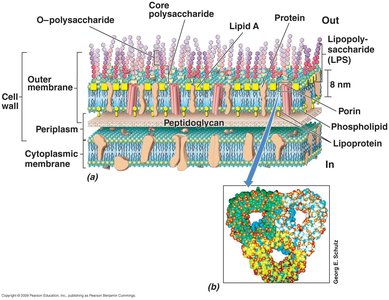
Peptidoglycan: A mesh-like polymer conferring rigidity and shape, and resisting turgor pressure.
Membranes: Inner and outer membranes regulate transport and protect cellular contents.
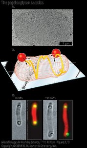
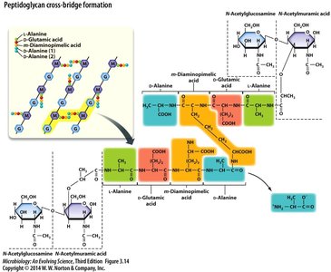
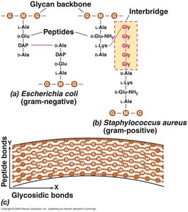
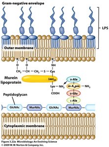
Peptidoglycan Structure and Function
Peptidoglycan, also known as murein, is a unique bacterial polymer composed of sugar chains cross-linked by peptides. It forms a sacculus that surrounds the cell, providing shape and resistance to osmotic pressure.
Sugar Chains: Alternating units of N-acetylglucosamine (NAG) and N-acetylmuramic acid (NAM).
Peptide Crosslinks: Short amino acid chains link the glycan strands, varying between Gram-positive and Gram-negative bacteria.
Role: Essential for cell division and a target for many antibiotics.
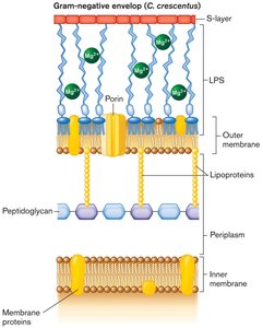
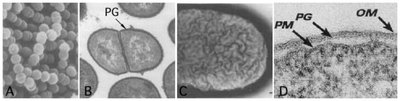
Gram-Positive vs. Gram-Negative Cell Walls
The cell wall structure differs significantly between Gram-positive and Gram-negative bacteria, affecting their staining properties and susceptibility to antibiotics.
Gram-Positive: Thick peptidoglycan layer, teichoic acids, no outer membrane.
Gram-Negative: Thin peptidoglycan layer, outer membrane with lipopolysaccharide (LPS), large periplasmic space.
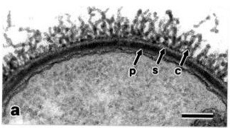
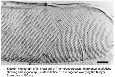
Surface Layers (S-Layers)
S-layers are crystalline protein arrays found on the surface of many bacteria and archaea, providing structural integrity and protection.
Composition: Made of protein or glycoprotein.
Function: Acts as a molecular sieve and protects against environmental stress.
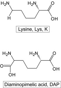
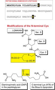
Molecular Structure of Peptidoglycan
Peptidoglycan cross-linking involves specific amino acids, such as diaminopimelic acid (DAP) and lysine, which differ between bacterial groups.
Cross-Bridge Formation: Peptide bonds link glycan chains, with DAP in Gram-negative and lysine in Gram-positive bacteria.
Importance: Provides mechanical strength and is a target for lysozyme and antibiotics.
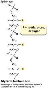
Teichoic Acids in Gram-Positive Bacteria
Teichoic acids are polymers found in the cell walls of Gram-positive bacteria, contributing to cell wall strength and ion regulation.
Structure: Glycerol or ribitol phosphate polymers with various side groups.
Function: Provides rigidity, regulates cation flow, and serves as a recognition site for bacteriophages.
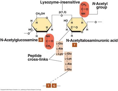
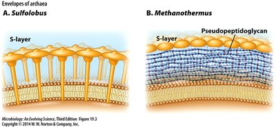
Cell Wall-Less Forms and Archaeal Cell Walls
Some bacteria, such as Mycoplasma, lack a cell wall and rely on tough membranes. Archaea possess unique cell wall structures, such as pseudomurein, which differ chemically from bacterial peptidoglycan.
Mycoplasma: Obligate intracellular parasites with pleomorphic shapes.
Pseudomurein: Contains N-talosaminuronic acid and β(1→3) glycosidic bonds, insensitive to lysozyme.
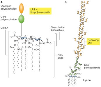
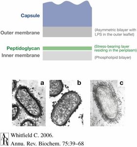
Gram-Negative Outer Membrane and Lipopolysaccharide (LPS)
The outer membrane of Gram-negative bacteria contains LPS, a complex molecule with important roles in pathogenicity and immune response.
Lipid A: Endotoxin component responsible for toxic effects.
Core Polysaccharide: Species-specific sugars, including KDO and heptose.
O-Antigen: Highly variable, used for serotyping.
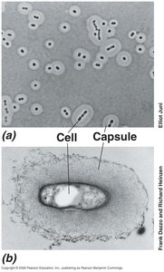
Capsules
Capsules are extracellular polysaccharide layers that provide additional protection and can be visualized using specific staining techniques.
Function: Protects against desiccation, phagocytosis, and environmental stress.
Composition: Highly variable among species.
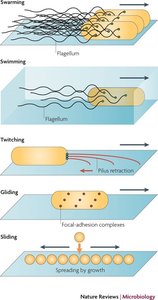
Motility
Surface Structures and Motility Mechanisms
Microbes employ various surface structures and mechanisms to move within their environments, including flagella, pili, and fimbriae.
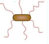
Flagella: Propeller-like appendages for swimming and swarming.
Pili and Fimbriae: Used for twitching and gliding motility.
Other Mechanisms: Gliding, sliding, and surfactant-mediated movement.
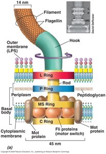
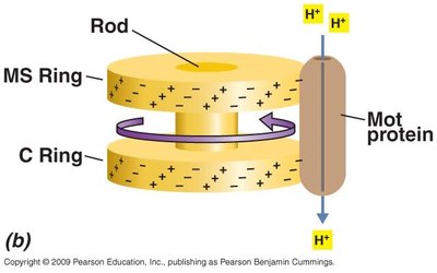
Flagella Structure and Function
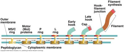
Flagella are complex structures that rotate to propel the cell. Their arrangement and assembly are key to microbial motility.
Arrangements: Polar, lophotrichous, peritrichous.
Components: Filament, hook, basal body, rings, and motor proteins.
Assembly: Sequential synthesis and insertion of structural components.
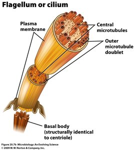
Eukaryotic Flagella
Eukaryotic flagella differ structurally and functionally from prokaryotic flagella, utilizing ATP-driven whip-like motion and microtubule-based architecture.
Structure: Central microtubules surrounded by outer doublets, enclosed by plasma membrane.
Function: Whip-like movement powered by ATP hydrolysis.
Summary Table: Gram-Positive vs. Gram-Negative Cell Envelope
Feature | Gram-Positive | Gram-Negative |
|---|---|---|
Peptidoglycan | Thick, heavily cross-linked | Thin, less cross-linked |
Teichoic Acids | Present | Absent |
Outer Membrane | Absent | Present (with LPS) |
Periplasm | Thin | Thick |
Capsule | Variable | Variable |
Conclusion
Microbial cell structure is highly diverse and adapted to environmental challenges. Protective layers, compartments, and motility appendages are essential for survival, pathogenicity, and ecological success. Understanding these features provides insight into microbial physiology and the basis for antimicrobial strategies.
