 Back
BackMicrobial Cell Structures: Prokaryotic and Eukaryotic Cells, Bacterial Cell Envelopes, and Specialized Structures
Study Guide - Smart Notes
 Tailored notes based on your materials, expanded with key definitions, examples, and context.
Microbial Cell Structures
Overview of Prokaryotic and Eukaryotic Cell Structures
Microorganisms are classified based on their cellular organization into prokaryotes and eukaryotes. Understanding the differences in their structures is fundamental to microbiology, as these differences influence physiology, genetics, and responses to antimicrobial agents.
Prokaryotes lack a true nucleus and membrane-bound organelles. Their genetic material is typically a single, circular double-stranded DNA molecule located in the nucleoid region.
Eukaryotes possess a nucleus surrounded by a nuclear membrane and various membrane-bound organelles such as mitochondria, endoplasmic reticulum, and Golgi apparatus.
Both cell types have a cell membrane, cytoplasm, and ribosomes, but differ in ribosome size and structure.
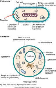
Key Differences Between Prokaryotic and Eukaryotic Cells
Nucleus: Prokaryotes have no nuclear membrane; eukaryotes have a true nucleus.
Chromosomes: Prokaryotes usually have a single, circular chromosome; eukaryotes have multiple, linear chromosomes.
Cell Wall: Most prokaryotes have a cell wall containing peptidoglycan; eukaryotic cell walls (if present) are chemically distinct (e.g., cellulose in plants, chitin in fungi).
Organelles: Eukaryotes have membrane-bound organelles; prokaryotes do not.
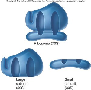
Ribosomes: Prokaryotic ribosomes are 70S (50S + 30S subunits); eukaryotic ribosomes are 80S (60S + 40S subunits).
Bacterial Cell Structures
External Structures
Bacteria possess a variety of external structures that contribute to motility, adherence, protection, and genetic exchange.
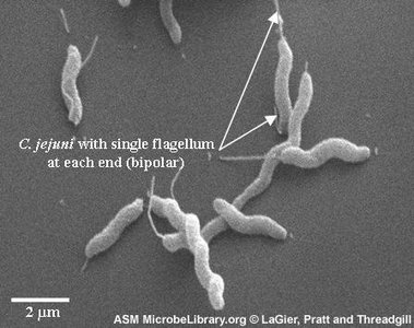
Flagella: Long, whip-like appendages used for motility. Structure includes the filament (flagellin protein), hook, and basal body (motor apparatus). Arrangements include monotrichous, lophotrichous, amphitrichous, and peritrichous.
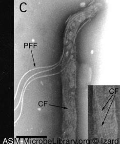
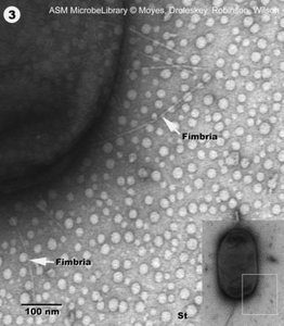
Pili (Fimbriae): Thin protein tubes, primarily in Gram-negative bacteria. Attachment pili (fimbriae) facilitate adherence; conjugation pili transfer DNA during conjugation.
Glycocalyx: Outer coating composed of polysaccharides and/or proteins. Exists as a capsule (organized, uniform) or slime layer (loose, variable). Functions include protection, biofilm formation, and evasion of host defenses.
Bacterial Cell Envelope
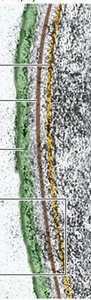
The cell envelope provides structural integrity and protection. It consists of the cell wall and cell membrane, with significant differences between Gram-positive and Gram-negative bacteria.
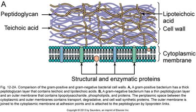
Cell Wall: Maintains shape, prevents osmotic lysis, and is composed of peptidoglycan (unique to bacteria).
Cell Membrane: Phospholipid bilayer with embedded proteins; functions in selective permeability, energy production, and biosynthesis.
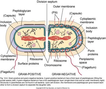
Gram-Positive vs. Gram-Negative Cell Walls
Gram-Positive: Thick peptidoglycan layer, teichoic acids, no outer membrane.
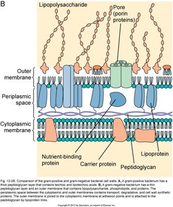
Gram-Negative: Thin peptidoglycan layer, outer membrane containing lipopolysaccharide (LPS), porin proteins, and a periplasmic space.
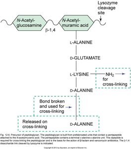
Peptidoglycan Structure and Synthesis
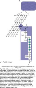
Peptidoglycan is a mesh-like polymer of sugars and amino acids, providing rigidity to the bacterial cell wall. It consists of repeating units of N-acetylglucosamine (NAG) and N-acetylmuramic acid (NAM) cross-linked by short peptides.
Enzymes called penicillin-binding proteins (PBPs) catalyze the cross-linking of peptidoglycan strands.
Lysozyme cleaves the β-1,4 linkage between NAG and NAM, compromising cell wall integrity.
Lipopolysaccharide (LPS) in Gram-Negative Bacteria
LPS is a major component of the outer membrane of Gram-negative bacteria. It consists of lipid A (endotoxin), a core polysaccharide, and an O-antigen. LPS contributes to structural integrity, protection from chemicals, and triggers strong immune responses in hosts.
Lipid A: Responsible for the toxic effects (endotoxin) when released during bacterial lysis.
O-antigen: Variable region used for serotyping.
Specialized Cell Envelopes
Acid-Fast Cell Walls: Contain mycolic acids, making them highly resistant to chemicals and dehydration. Basis for the acid-fast stain (e.g., Mycobacterium).
Bacteria Without Cell Walls: Some bacteria (e.g., Mycoplasma) lack cell walls but have membranes stabilized by sterols, resulting in pleomorphism.
Bacterial Internal Structures
Cytoplasm: Gel-like matrix composed of water, nutrients, and cell components.
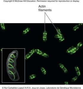
Cytoskeleton: Protein filaments (actin-like, tubulin-like) involved in cell shape, division, and intracellular transport.
Chromosome: Usually a single, circular, double-stranded DNA molecule located in the nucleoid.
Plasmids: Small, circular DNA molecules that replicate independently and may carry genes for antibiotic resistance or virulence.
Ribosomes: Sites of protein synthesis; prokaryotic ribosomes are 70S (50S + 30S subunits).
Inclusion Bodies: Storage granules for nutrients and other substances.
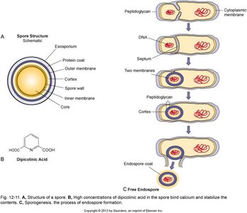
Endospores
Endospores are highly resistant, dormant structures formed by certain Gram-positive bacteria (e.g., Bacillus, Clostridium) in response to adverse conditions. They allow survival through extreme heat, desiccation, chemicals, and radiation.
Life Cycle: Includes a vegetative cell (active, growing) and an endospore (dormant, resistant).
Sporulation: Process of endospore formation (not reproduction).
Germination: Return to vegetative growth when conditions improve.
Summary Table: Comparison of Prokaryotic and Eukaryotic Cell Structures
Feature | Prokaryotes | Eukaryotes |
|---|---|---|
Nucleus | Absent | Present |
Chromosomes | Single, circular | Multiple, linear |
Cell Wall | Peptidoglycan (most) | Cellulose, chitin, or absent |
Organelles | Absent | Present |
Ribosomes | 70S | 80S |
Flagella | Simple, flagellin-based | Complex, microtubule-based |
Additional info: Some bacteria have sterols in their membranes, and many eukaryotes (plants, algae, fungi) have cell walls and may possess plasmids. The presence and arrangement of external structures such as flagella and pili are important for motility, adherence, and genetic exchange. The Gram stain is a critical diagnostic tool for distinguishing bacterial types based on cell wall structure.
