 Back
Microbial Cell Surface Structures, Inclusions, Endospores, and Locomotion
Study Guide - Smart Notes
 Tailored notes based on your materials, expanded with key definitions, examples, and context.
Basic Cell Structures
Prokaryotic vs. Eukaryotic Cell Structure
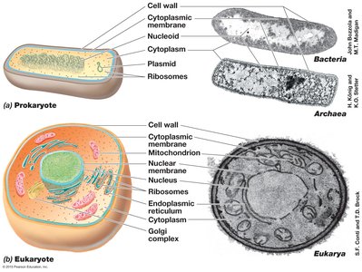
Microbial cells are classified as prokaryotes (Bacteria and Archaea) or eukaryotes (Eukarya), each with distinct structural features. Understanding these differences is fundamental to microbiology.
Prokaryotes lack a membrane-bound nucleus and organelles; their genetic material is located in the nucleoid.
Eukaryotes possess a nucleus and various organelles such as mitochondria, endoplasmic reticulum, and Golgi complex.
Both cell types have a cytoplasmic membrane, ribosomes, and cytoplasm.
Bacteria and Archaea differ in cell wall composition and other structural features.
Cell Surface Structures
Capsules
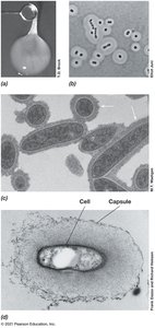
Capsules are extracellular layers composed of polysaccharides and/or proteins that surround the cell wall in many bacteria. They play critical roles in microbial survival and pathogenicity.
Capsules prevent penetration of particles and compounds, aiding in protection.
They facilitate attachment to surfaces and host tissues.
Capsules help microbes evade the immune system and prevent desiccation.
Capsules are easily viewed under a light microscope.
Examples: Leuconostoc mesenteroides (thick dextran slime layer), Acinetobacter, Rhodobacter, Rhizobium.
Slime Layers
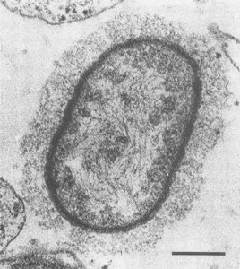
Slime layers are similar to capsules but are more easily disrupted and harder to visualize. They also consist of polysaccharides and/or proteins and serve similar protective and adhesive functions.
Slime layers are less tightly bound to the cell than capsules.
They aid in attachment and protection but are more susceptible to environmental disruption.
Fimbrae
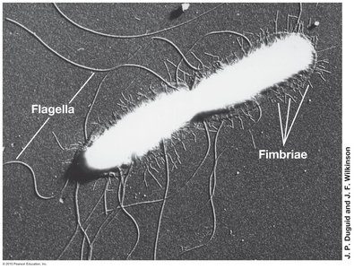
Fimbrae are short, filamentous protein structures present in large numbers on the surface of many bacteria. They are essential for attachment and biofilm formation.
Hundreds to thousands per cell.
Facilitate attachment to surfaces and host tissues.
Play a role in pathogenesis for species such as Salmonella, Neisseria gonorrhoeae, and Bordetella pertussis.
Pili
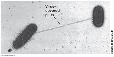
Pili are longer and less numerous than fimbrae. They are involved in genetic exchange and motility.
Sex pili facilitate conjugation (genetic exchange between bacteria).
Type IV pili assist in cell adhesion and twitching motility.
Pathogens such as Vibrio cholerae and Neisseria gonorrhoeae use pili for locating attachment sites.
Cell Inclusions
Carbon Storage Inclusions
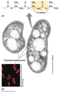
Microbes store excess carbon in specialized inclusions, such as poly-β-hydroxyalkanoates (PHAs) and glycogen. These inclusions help maintain osmotic balance and provide energy reserves.
PHAs are synthesized when carbon is abundant.
Glycogen is used by some organisms for carbon storage.
These inclusions are visible as large granules within cells.
Polyphosphate and Sulfur Granules
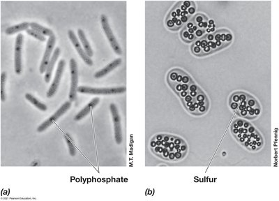
Microbes can store essential elements such as phosphorus and sulfur in granules for later use.
Polyphosphate granules store phosphorus, a frequently limiting nutrient.
Elemental sulfur granules are stored by sulfur-oxidizing bacteria for energy when hydrogen sulfide is unavailable.
Magnetic Inclusions (Magnetosomes)
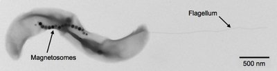
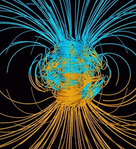
Magnetosomes are intracellular structures composed of magnetite or greigite, allowing bacteria to orient themselves along Earth's magnetic field.
Found in anaerobic bacteria, not in Archaea.
Guide bacteria to low oxygen environments.
Vary in shape: square, rectangular, or spike-shaped.
Gas Vesicles
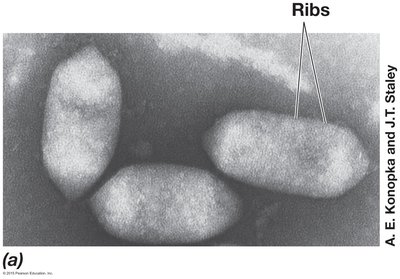
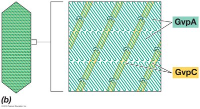
Gas vesicles are protein-bound structures that provide buoyancy to planktonic bacteria, allowing them to position themselves optimally in the water column.
Permeable only to gases.
Composed of GvpA (forms ribs) and GvpC (strengthens vesicle).
Not found in microbial Eukarya.
Unusual Morphologies
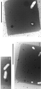
Bacteria and Archaea exhibit a wide range of unusual cell shapes, such as the square-shaped Haloquadratum.
Cell shape diversity reflects adaptation to specific environments.
Gas Vacuoles in Cyanobacteria
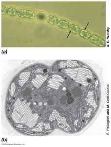
Cyanobacteria possess clusters of gas vesicles called gas vacuoles, which help them regulate buoyancy.
Gas vacuoles are visible as clear areas within cells.
Examples: Anabaena and Microcystis.
Endospores
Endospore Formation and Function
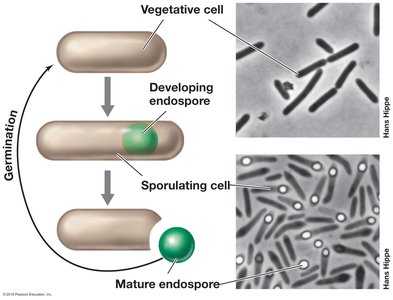
Endospores are highly resilient survival structures produced by certain gram-positive bacteria in response to environmental stress, such as starvation.
Endospores exhibit very low metabolic activity.
They are not found in Archaea, suggesting evolutionary development after the bacterial split.
Endospores allow bacteria to survive extreme conditions.
Botulism and Endospores
Botulism is caused by the neurotoxin produced by Clostridium botulinum, a bacterium capable of sporulation. The toxin is destroyed by heating above 80°C or exposure to low pH.
Botulism is a severe or fatal food poisoning.
Most cases are infant, wound, or foodborne (often from canned foods).
Microbial Locomotion
Flagella and Types of Flagellation
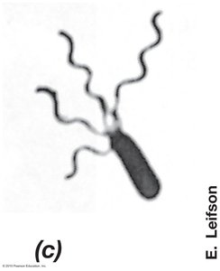
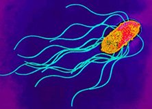
Flagella are whip-like appendages that enable motility in many bacteria. The arrangement and number of flagella vary among species.
Polar flagellation: Flagella attached at one or both ends of the cell.
Monotrichous: Single flagellum at one pole.
Lophotrichous: Multiple flagella at one pole.
Amphitrichous: Multiple flagella at both poles.
Peritrichous flagellation: Flagella distributed over the entire cell surface.
Flagellar Structure and Function
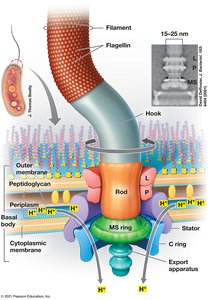
The bacterial flagellum is a complex structure composed of several protein rings and a filament. Its rotation is powered by the flow of protons across the membrane.
Flagellin protein sequence is highly conserved, indicating early evolutionary origin.
Flagellum rotates like a propeller.
Key components: hook, L-ring, P-ring, MS-ring, C-ring, Mot proteins (stator).
Flagellar Rotation Mechanism
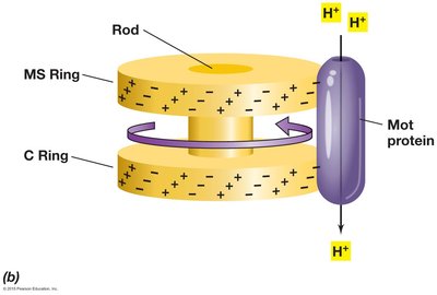
Flagellar rotation is driven by the Mot complex, which allows protons to flow back into the cytoplasm, generating torque.
About 1000 protons are translocated per rotation.
Proton flow attracts negative charges on MS- and C-rings, causing rotation.
Flagella Rate of Rotation
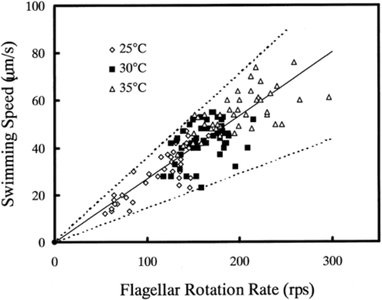
Bacterial flagella can rotate at high speeds, resulting in rapid cell movement. The rate of rotation and swimming speed are influenced by environmental conditions such as temperature.
Some bacteria move at speeds up to 60 cell lengths per second.
Swimming speed correlates with flagellar rotation rate.
Flagellar Movement Patterns
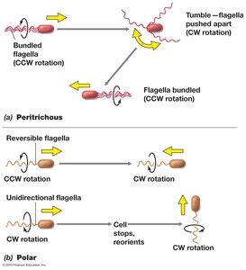
Bacteria exhibit different movement patterns depending on flagellar arrangement.
Peritrichous: "Bundle and tumble"—counterclockwise (CCW) rotation moves forward, clockwise (CW) causes tumbling.
Polar: Reversible flagella (CCW forward, CW backward) or unidirectional flagella (CW forward with reorientation).
Gliding Motility
Gliding motility is a slower form of movement requiring contact with a solid surface. It is observed in certain bacteria and involves various mechanisms.
Mechanisms include polysaccharide slime secretion, twitching motility via type IV pili, and ratcheting mechanisms.
Gliding allows exploitation of new resources and social behaviors.
No gliding Archaea are known.
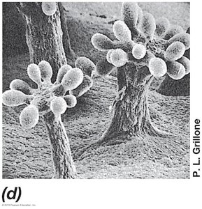
Taxis: Directed Movement
Taxis refers to the directed movement of microbes in response to environmental stimuli. Chemotaxis, aerotaxis, phototaxis, and other forms of taxis are crucial for microbial survival and adaptation.
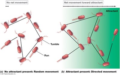
Chemotaxis: Movement toward or away from chemicals, sensed by membrane-bound chemoreceptors.
Cells respond to temporal, not spatial, changes in chemical concentration.
Without a gradient, movement is random; with a gradient, movement is directed.
Measuring Chemotaxis
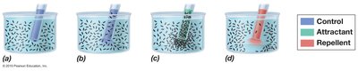
Chemotaxis can be measured using a capillary tube assay, where attractants or repellents are placed in a tube and cell concentrations are compared to controls.
Cells accumulate in the tube if attracted, or avoid it if repelled.
Aerotaxis and Phototaxis
Aerotaxis is movement toward or away from oxygen, while phototaxis is movement in response to light. Both are mediated by similar proteins as chemotaxis.
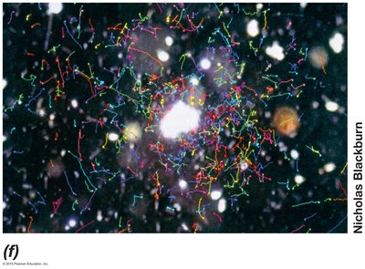
Aerotactic bacteria move toward oxygen-producing phototrophs.
Phototactic bacteria move toward higher light intensities, often for photosynthesis.
Other Forms of Taxis
Microbes also exhibit osmotaxis (response to ionic strength) and hydrotaxis (response to water availability), important for survival in diverse environments.
Osmotaxis: Movement toward areas of high ionic strength.
Hydrotaxis: Movement toward water, as seen in desert cyanobacteria.
Additional info: These notes expand on brief points with academic context, definitions, and examples to provide a comprehensive study guide for microbiology students.
