 Back
BackMicrobial Culture Transfer Techniques and Serratia marcescens
Study Guide - Smart Notes
 Tailored notes based on your materials, expanded with key definitions, examples, and context.
Microbial Culture Transfer Techniques
Principles of Microbial Cultures
Microbial cultures are populations of microorganisms grown in controlled environments, either in liquid (broth) or solid media. These cultures are essential for studying microbial physiology, genetics, and pathogenicity.
Broth cultures: Liquid media used for growing microorganisms in suspension.
Solid media: Agar-based media used for isolating colonies and observing growth patterns.
Slants: Solid media set at an angle in test tubes, providing a larger surface area for growth.
Deeps: Solid media in upright tubes, used for studying motility and oxygen requirements.
Inoculation Tools and Techniques
The transfer of microorganisms between media types is performed using specialized tools and aseptic techniques to prevent contamination.
Inoculating loop: A wire loop used to transfer microorganisms, suitable for broth, slant, and streaking on plates.
Inoculating needle: A straight wire used for stabbing cultures into deep agar tubes.
Aseptic technique: Procedures such as flaming tools and tube necks to maintain sterility during transfers.
Step-by-Step Transfer Procedure
Proper transfer technique is crucial for reliable results in microbiology experiments.
Label test tubes with masking tape for identification.
Secure tubes in the palm, forming a V shape for stability.
Flame the inoculating loop or needle to sterilize.
Aseptically uncap the tubes and flame the necks to prevent contamination.
Retrieve a loopful of culture from the source tube.
Transfer the culture to the fresh media (broth, slant, or deep).
Flame the necks again, recap tubes, and re-flame the loop or needle.
Types of Transfers and Incubation Conditions
Different transfer techniques are used depending on the experimental goals and media type.
Transfer from slant to broth (incubate at 40°C and room temperature).
Transfer from slant to slant (incubate at 40°C and room temperature).
Transfer from slant to deep using a needle (incubate at 40°C and room temperature).
Example: After inoculation, one set of tubes (broth, slant, deep) is incubated at room temperature, and another at 40°C to observe differences in microbial growth.
Serratia marcescens: Characteristics and Growth
Overview of Serratia marcescens
Serratia marcescens is a Gram-negative rod-shaped bacterium commonly found in clinical, environmental, and animal sources.
Gram-negative rods: Cell wall structure lacks thick peptidoglycan, stains pink in Gram stain.
Size: 0.5 - 0.8 x 0.9 – 2.0 µm.
Motility: Motile with peritrichous flagella (flagella distributed over the entire cell surface).
Metabolism: Facultative anaerobe, capable of both respiratory and fermentative metabolism.
Growth temperature: Optimal growth between 30°C and 37°C, but can grow at room temperature and 40°C.
Habitat: Found in human clinical specimens, soil, water, plant surfaces, digestive tracts of rodents and insects.
Growth Patterns in Different Conditions
Serratia marcescens exhibits distinct growth characteristics depending on incubation temperature and media type.
At room temperature, S. marcescens often produces a characteristic red pigment (prodigiosin).
At higher temperatures (e.g., 40°C), pigment production may decrease or be absent.
Growth in slants and deeps can be used to study motility and oxygen requirements.
Example: Comparing S. marcescens grown at room temperature and 40°C demonstrates temperature-dependent pigment production. 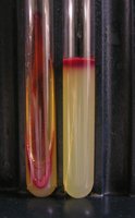
Applications and Importance
Serratia marcescens is used in laboratory studies for its pigment production and as a model organism for microbial transfer techniques.
Demonstrates principles of microbial growth and metabolism.
Used in teaching aseptic technique and culture transfer methods.
Clinical relevance: Can cause opportunistic infections in humans.
Additional info: The image included shows slant and deep tubes inoculated with Serratia marcescens, highlighting the red pigment produced at room temperature, which is a classic demonstration in microbiology labs for observing microbial growth and pigment production.
