Back
BackMicrobial Diseases of the Nervous System: Structure, Pathogenesis, and Clinical Features
Study Guide - Smart Notes
 Tailored notes based on your materials, expanded with key definitions, examples, and context.
Microbial Diseases of the Nervous System
Function and Protection of the Nervous System
The nervous system is essential for maintaining coordination and homeostasis in the body. It transmits nerve impulses rapidly, resulting in quick but less prolonged responses compared to other systems. The central nervous system (CNS) is protected from injury and infection by bone (skull and vertebrae) and specialized structures such as the blood-brain barrier, which restricts pathogen entry. Diseases affecting the CNS can cause severe consequences, including deafness, blindness, learning disabilities, paralysis, and death.
Blood-brain barrier: A selective barrier that prevents most pathogens in the bloodstream from entering the brain and spinal cord.
Cerebrospinal fluid (CSF): Lacks many immune defenses found in blood, making the CNS vulnerable if pathogens breach protective barriers.
Trauma: Disruption of CNS defenses can allow pathogens to invade.
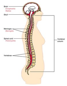
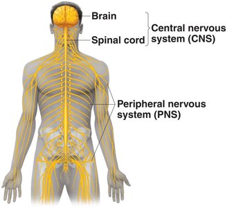
Anatomy and Physiology Review
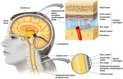
The CNS consists of the brain and spinal cord, protected by the skull and vertebral column. The meninges are three protective membranes (dura mater, arachnoid mater, pia mater) surrounding the CNS, and the CSF circulates within these layers, providing cushioning and nutrient transport.
Meninges: Dura mater (outer), arachnoid mater (middle), pia mater (inner).
CSF: Found in the subarachnoid space; acts as a shock absorber and medium for exchange.
Bacterial Diseases of the Nervous System
Bacterial Meningitis
Meningitis is the inflammation of the meninges, which can be caused by bacteria, viruses, fungi, or protozoa. Bacterial meningitis is more severe than viral, often resulting in brain damage or death. The three major bacterial causes are Haemophilus influenzae, Neisseria meningitidis, and Streptococcus pneumoniae. These bacteria are protected by capsules and can rapidly replicate in the bloodstream, gaining access to the CSF.
Symptoms: Fever, headache, stiff neck, nausea, vomiting, convulsions, coma, shock, and death.
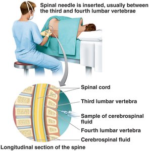
Diagnosis: Gram stain or latex agglutination of CSF; lumbar puncture (spinal tap) is used to collect CSF.
Treatment: Cephalosporins are administered before pathogen identification.
Haemophilus influenzae Meningitis
Gram-negative, pleomorphic coccobacilli with a capsule.
Requires blood factors for growth; part of normal throat microbiota.
Primarily affects children under age 4, especially around 6 months.
Can also cause pneumonia, otitis media, and epiglottitis.
Neisseria Meningitidis (Meningococcal Meningitis)
Gram-negative cocci with a capsule; many healthy carriers.
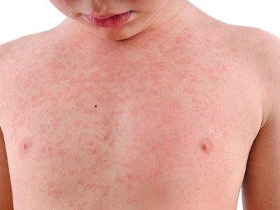
Begins as throat infection, followed by a characteristic rash and rapid progression to death.
Symptoms caused by endotoxin; most cases in children under 2 years.
Survivors may experience deafness; antibiotics reduce mortality to 9–12%.
Pneumococcal Meningitis (Streptococcus pneumoniae)
Gram-positive diplococci; associated with pneumonia and septicemia.
70% of people are healthy carriers; most common in children (1 month to 4 years).
Mortality: 30% in children, 80% in elderly; increasing antibiotic resistance.
Viral Meningitis
Viral meningitis is more common and milder than bacterial meningitis. It is usually caused by enteroviruses, which grow well in the throat and intestinal tract. It can also be a complication of mumps, chickenpox, and influenza.
Diagnosis: Lumbar puncture to collect CSF.
Listeriosis (Listeria monocytogenes)
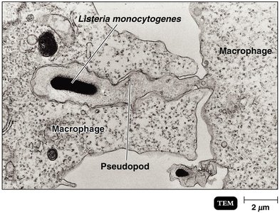
Listeria monocytogenes is a Gram-positive organism that reproduces in phagocytes. It is a psychrophile, often causing foodborne infections. Listeriosis may be asymptomatic or mild in healthy adults but can cause meningitis in newborns, immunosuppressed individuals, pregnant women, and cancer patients. It can cross the placenta, causing spontaneous abortion or stillbirth.
Transmission: Ingestion of contaminated food.
Pathogenesis: Reproduces in phagocytes, evading immune response.
Tetanus (Lockjaw)
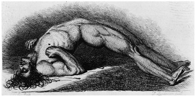
Tetanus is caused by Clostridium tetani, a Gram-positive, endospore-forming, obligate anaerobe. It grows in deep wounds and releases tetanospasmin, a neurotoxin that blocks muscle relaxation, causing muscle spasms and rigidity.
Prevention: Vaccination with tetanus toxoid (DTaP) and booster (dT).
Treatment: Tetanus immune globulin.
Botulism
Botulism is caused by Clostridium botulinum, a Gram-positive, endospore-forming, obligate anaerobe. Intoxication occurs from ingesting botulinal toxin, which blocks neurotransmitter release, causing flaccid paralysis.
Prevention: Proper canning, nitrites in sausages to prevent endospore germination.
Treatment: Supportive care and antitoxin.
Types: Type A (60–70% fatality), Type B (25% fatality), Type E (marine/lake sediments).
Leprosy (Hansen’s Disease)
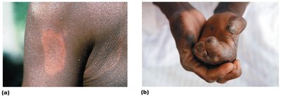
Leprosy is caused by Mycobacterium leprae, an acid-fast rod that grows best at 30°C in peripheral nerves and skin cells. Transmission requires prolonged contact with an infected person. The disease manifests in two forms: tuberculoid (neural) and lepromatous (progressive).
Tuberculoid form: Loss of sensation in skin areas; positive lepromin test (cell-mediated immunity).
Lepromatous form: Disfiguring nodules; negative lepromin test (lack of immunity).
Viral Diseases of the Nervous System
Poliomyelitis (Infantile Paralysis)
Poliomyelitis is caused by poliovirus (an enterovirus of the Picornaviridae family). It is transmitted by ingestion and has three strains. Most cases are asymptomatic, but in rare cases, the virus enters the CNS and selectively destroys motor neurons, causing paralysis.
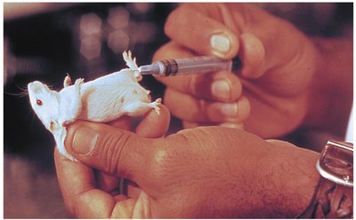
Prevention: Vaccination (Salk vaccine - inactivated, Sabin vaccine - live attenuated, and newer IPV).
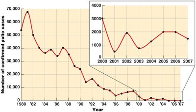
Incidence: Dramatic reduction in polio cases worldwide due to vaccination.
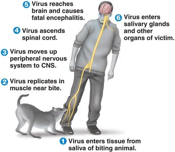
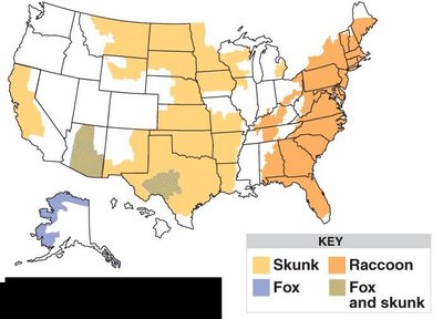
Rabies
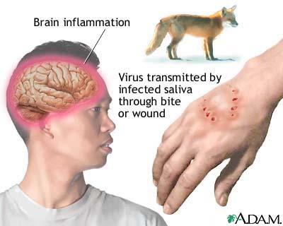
Rabies is caused by the rabies virus (Lyssavirus, family Rhabdoviridae), a single-stranded RNA virus. It is a zoonosis, transmitted from the saliva of rabid animals via bites. The virus multiplies in skeletal muscles, then travels retrograde to the CNS, causing encephalitis and eventually spreading to salivary glands and other organs.
Symptoms: Muscle spasms of the mouth and pharynx, hydrophobia, behavioral changes.
Forms: Furious rabies (restlessness, excitability), paralytic rabies (unawareness).
Treatment: Preexposure prophylaxis (HDCV vaccine), postexposure prophylaxis (HDCV vaccine and rabies immune globulin).
Prevention: Vaccination of pets and, if necessary, wild populations.
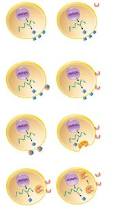
Prion Diseases
Prions and Spongiform Encephalopathies
Prions are proteinaceous infectious particles that convert normal proteins into abnormal forms, leading to neurodegenerative diseases that are chronic and fatal. Prion diseases are inherited or transmissible by ingestion, transplant, or contaminated surgical instruments. Examples include sheep scrapie, Creutzfeldt-Jakob disease, Gerstmann-Sträussler-Scheinker syndrome, fatal familial insomnia, and mad cow disease.
Cellular Prion Protein (PrPC): Functions in cell adherence and survival.
Scrapie Protein (PrPSc): Accumulates in brain cells, forming plaques and causing cell death.
Prevention: Surgical instruments sterilized by NaOH and extended autoclaving at 134°C.
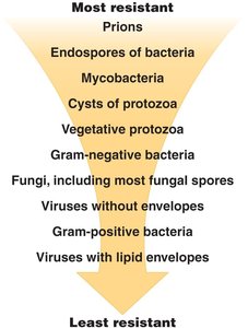
Resistance of Microbial Agents
Prions are among the most resistant infectious agents, followed by endospores of bacteria, mycobacteria, and cysts of protozoa. The resistance decreases through vegetative protozoa, gram-negative bacteria, fungi, viruses, and gram-positive bacteria, with viruses with lipid envelopes being the least resistant.
Most Resistant | Least Resistant |
|---|---|
Prions | Viruses with lipid envelopes |
Endospores of bacteria | Gram-positive bacteria |
Mycobacteria | Viruses without envelopes |
Cysts of protozoa | Fungi, including most fungal spores |
Vegetative protozoa | Gram-negative bacteria |