 Back
BackMicrobial Diseases of the Respiratory System: Structure, Pathogenesis, and Clinical Syndromes
Study Guide - Smart Notes
 Tailored notes based on your materials, expanded with key definitions, examples, and context.
Microbial Diseases of the Respiratory System
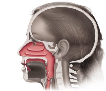
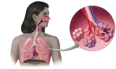
Overview of the Respiratory System
The respiratory system is divided into the upper and lower respiratory tracts, each with distinct anatomical structures and associated microbiota. Understanding these divisions is essential for recognizing the pathogenesis and clinical presentation of respiratory infections.
Upper respiratory system: Includes the nasal cavity, sinuses, pharynx, larynx, and associated structures.
Lower respiratory system: Comprises the trachea, bronchi, bronchioles, lungs, and alveoli.
Normal Microbiota of the Respiratory System
The upper respiratory tract contains a diverse microbiota that suppresses pathogens through competitive inhibition. In contrast, the lower respiratory tract is typically sterile due to the action of the mucociliary escalator and immune defenses.
Competitive inhibition: Normal flora outcompete potential pathogens for nutrients and attachment sites.
Sterility of lower tract: Maintained by physical and immunological barriers.
Diseases of the Upper Respiratory System
Common Upper Respiratory Infections
Several diseases affect the upper respiratory tract, often presenting with sore throat, inflammation, and sometimes systemic symptoms.
Pharyngitis: Inflammation of the pharynx.
Laryngitis: Inflammation of the larynx.
Tonsillitis: Inflammation of the tonsils.
Sinusitis: Inflammation of the sinuses.
Epiglottitis: Inflammation of the epiglottis, often caused by Haemophilus influenzae type b.
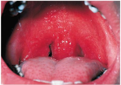
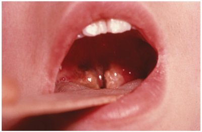
Streptococcal Pharyngitis (Strep Throat)
Strep throat is a common bacterial infection of the throat caused by Streptococcus pyogenes. It is characterized by resistance to phagocytosis and the production of toxins.
Pathogen: Streptococcus pyogenes (Group A Streptococcus).
Virulence factors: Streptokinases (lyse clots), streptolysins (cytotoxic), and resistance to phagocytosis.
Diagnosis: Enzyme immunoassay (EIA) tests.
Complication: Scarlet fever, caused by erythrogenic toxin produced by lysogenized strains.
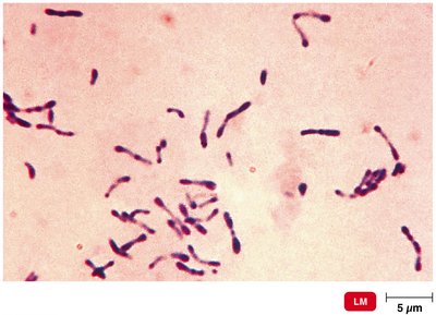
Diphtheria
Diphtheria is a serious upper respiratory infection caused by Corynebacterium diphtheriae, a gram-positive rod. The disease is characterized by the formation of a tough, grayish membrane in the throat and the production of a potent exotoxin.
Pathogen: Corynebacterium diphtheriae.
Toxin: Diphtheria toxin (produced by lysogenized bacteria).
Prevention: DTaP vaccine (diphtheria toxoid).
The Common Cold
The common cold is a viral infection of the upper respiratory tract, most frequently caused by rhinoviruses and coronaviruses.
Major causative agents: Rhinoviruses (30–50%), coronaviruses (10–15%).
Symptoms: Runny nose, sore throat, cough, mild fever.
Diseases of the Lower Respiratory System
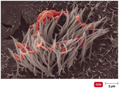
Pertussis (Whooping Cough)
Pertussis is a highly contagious bacterial disease caused by Bordetella pertussis. It is characterized by severe coughing fits and is preventable by vaccination.
Pathogen: Bordetella pertussis (gram-negative coccobacillus).
Virulence factors: Capsule, tracheal cytotoxin (damages ciliated cells), pertussis toxin.
Prevention: DTaP vaccine (acellular pertussis components).
Stages:
Catarrhal stage: mild, cold-like symptoms.
Paroxysmal stage: severe, violent coughing fits.
Convalescence stage: gradual recovery.
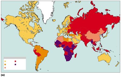
Tuberculosis (TB)
Tuberculosis is a chronic infectious disease primarily affecting the lungs, caused by Mycobacterium tuberculosis. It is transmitted via airborne droplets and can become latent or progress to active disease.
Pathogen: Mycobacterium tuberculosis (acid-fast rod).
Transmission: Human-to-human via respiratory droplets.
Other species: M. bovis (rare in humans), M. avium-intracellulare (in immunocompromised patients).
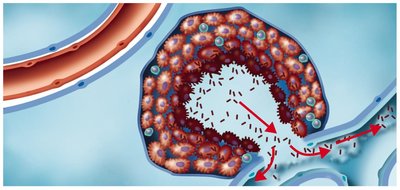
Pathogenesis of Tuberculosis
The pathogenesis of TB involves several stages, from initial infection to possible dissemination throughout the body.
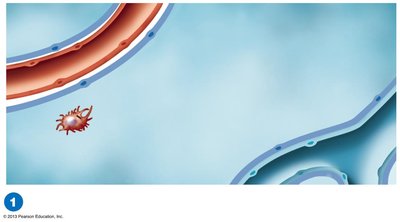
Tubercle bacilli reach the alveoli and are ingested by macrophages; some survive and establish infection without symptoms.
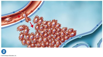
Bacilli multiply, attracting more macrophages and forming an early tubercle; inflammation damages lung tissue.
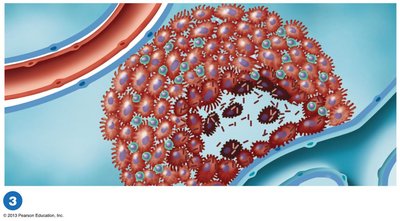
Caseous center forms as macrophages die; bacilli may remain dormant (latent TB) or lesions may calcify.
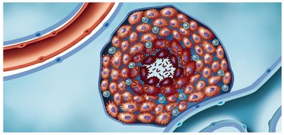
In some, the caseous center liquefies, forming a tuberculous cavity where bacilli multiply.
Rupture of the tubercle releases bacilli into bronchioles, spreading infection.
Diagnosis and Treatment of Tuberculosis
Diagnosis: Tuberculin skin test, chest X-ray or CT, acid-fast staining, bacterial culture.
Treatment: Prolonged therapy with multiple antibiotics.
Prevention: BCG vaccine (live, avirulent M. bovis), not widely used in the U.S.
Pneumonias of Bacterial Origin
Pneumococcal Pneumonia
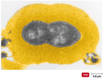
Caused by Streptococcus pneumoniae, this is the most common type of bacterial pneumonia. It is characterized by fluid-filled alveoli and impaired oxygen exchange.
Pathogen: Streptococcus pneumoniae (encapsulated diplococci).
Symptoms: Cough, fever, chest pain, difficulty breathing.
Diagnosis: Serological typing.
Treatment: Macrolides, fluoroquinolones.
Prevention: Pneumococcal vaccine.
Haemophilus influenzae Pneumonia
This pneumonia is caused by Haemophilus influenzae, a gram-negative coccobacillus. It often affects individuals with underlying health conditions.
Predisposing factors: Alcoholism, poor nutrition, cancer, diabetes.
Symptoms: Similar to pneumococcal pneumonia.
Diagnosis: Isolation on special media.
Treatment: Cephalosporins.
Mycoplasmal Pneumonia (Walking Pneumonia)
Caused by Mycoplasma pneumoniae, this form of pneumonia is generally mild and common in children and young adults.
Pathogen: Mycoplasma pneumoniae (pleomorphic, wall-less bacteria).
Symptoms: Mild but persistent cough, low fever, headache.
Diagnosis: PCR and serological testing.
Treatment: Tetracyclines.
Legionellosis (Legionnaires' Disease)
Legionellosis is caused by Legionella pneumophila, a gram-negative rod found in water. It is transmitted by inhaling aerosols and can cause severe pneumonia, especially in older adults.
Symptoms: Severe pneumonia, often in older men who smoke or drink heavily.
Diagnosis: Culture on selective media, DNA probe.
Treatment: Erythromycin.
Chlamydial Pneumonia
Caused by Chlamydophila pneumoniae, this pneumonia is typically mild and common among young people.
Transmission: Human-to-human.
Symptoms: Mild respiratory illness.
Diagnosis: Serological tests.
Treatment: Tetracyclines.
Viral and Fungal Respiratory Diseases
Viral Pneumonia
Viral pneumonia often occurs as a complication of other viral infections such as influenza, measles, or chickenpox. It is suspected when no bacterial cause is identified.
Respiratory Syncytial Virus (RSV)
RSV is a major cause of pneumonia in infants, leading to cell fusion (syncytium formation) in cell culture.
Symptoms: Pneumonia in infants.
Diagnosis: Serological tests for virus and antibodies.
Treatment: Ribavirin, palivizumab.
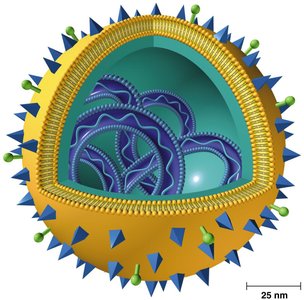
Influenza (Flu)
Influenza is a highly contagious viral disease characterized by fever, chills, headache, and muscle aches. The virus undergoes frequent antigenic changes, complicating immunity and vaccine development.
Structure: Hemagglutinin (HA) spikes for attachment, neuraminidase (NA) spikes for release.
Antigenic shift: Major changes due to genetic recombination between strains.
Antigenic drift: Minor changes due to point mutations.
Treatment: Zanamivir and oseltamivir (neuraminidase inhibitors).
Prevention: Multivalent vaccine.
Fungal Respiratory Diseases
Fungi can cause systemic respiratory infections, particularly in immunocompromised individuals or those with underlying conditions such as cancer or diabetes.
Common pathogens: Aspergillus fumigatus, Mucor, Rhizopus.
Predisposing factors: Immunocompromised state, cancer, diabetes.
Additional info: This guide covers the major microbial diseases of the respiratory system, including their causative agents, pathogenesis, clinical features, diagnosis, and prevention. It is suitable for exam preparation in a college-level microbiology course.
