 Back
BackMicrobial Diseases of the Respiratory System: Structure, Pathogenesis, and Clinical Features
Study Guide - Smart Notes
 Tailored notes based on your materials, expanded with key definitions, examples, and context.
Diseases of the Respiratory System
Overview of the Respiratory System
The respiratory system is divided into the upper and lower respiratory tracts, each with distinct anatomical structures and associated microbiota. Understanding these divisions is essential for recognizing the pathogenesis and clinical presentation of respiratory infections.
Upper respiratory system: Includes the nasal cavity, pharynx, larynx, sinuses, middle ear, and associated structures.
Lower respiratory system: Comprises the trachea, bronchi, bronchioles, alveoli, and lungs.
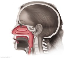
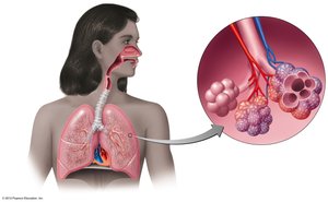
Normal Microbiota of the Respiratory System
The upper respiratory tract harbors a diverse normal flora that suppresses pathogens through competitive inhibition. The lower respiratory tract is typically sterile due to the action of alveolar macrophages and mucociliary clearance.
Common normal flora: Diphtheroids, Staphylococci, Micrococci, Bacillus, Streptococcus pneumoniae, Haemophilus influenzae, Neisseria meningitidis
Defense mechanisms: Mucociliary escalator, alveolar macrophages
Upper Respiratory System Diseases
General Infections
Diseases of the upper respiratory tract include pharyngitis, laryngitis, tonsillitis, sinusitis, and epiglottitis. These conditions are caused by a variety of bacterial and viral pathogens and can lead to complications if untreated.
Streptococcal Pharyngitis (Strep Throat)
Streptococcal pharyngitis is a common bacterial infection of the throat caused by Streptococcus pyogenes (Group A, beta-hemolytic). It is characterized by local inflammation, fever, and sometimes tonsillitis or otitis media.
Virulence factors: Resistance to phagocytosis, streptokinases (lyse clots), streptolysins (cytotoxic)
Diagnosis: Enzyme immunoassay (EIA), blood agar culture, bacitracin sensitivity
Treatment: Penicillin
Complications: Scarlet fever (erythrogenic toxin), rheumatic fever
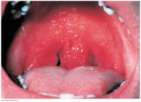
Scarlet Fever
Scarlet fever is a complication of strep throat caused by erythrogenic toxin produced by lysogenized S. pyogenes. It presents with a pink-red rash, high fever, and a characteristic 'strawberry tongue.'
Diagnosis: Dick test (skin test for immunity)
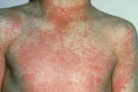

Diphtheria
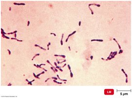
Diphtheria is caused by Corynebacterium diphtheriae, a Gram-positive, non-endospore-forming, club-shaped rod. The disease is mediated by a potent exotoxin that inhibits protein synthesis, leading to local and systemic effects.
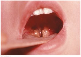
Symptoms: Sore throat, fever, malaise, neck swelling, gray membrane in throat (may block airway)
Transmission: Airborne droplets
Treatment: Antibiotics (penicillin, erythromycin) and antitoxins
Prevention: DTaP vaccine (diphtheria toxoid)
Diagnosis: Loeffler slant, Tellurite agar, demonstration of toxigenicity
Otitis Media
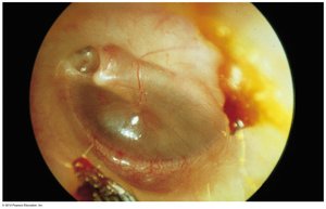
Otitis media is a middle ear infection, commonly caused by Streptococcus pneumoniae, Haemophilus influenzae, Moraxella catarrhalis, S. pyogenes, and S. aureus. It is characterized by ear pain and a bulging eardrum.
Incidence: Reduced by pneumococcal vaccine
The Common Cold
The common cold is primarily caused by rhinoviruses and coronaviruses. It is highly contagious and presents with mild upper respiratory symptoms.
Symptoms: Runny nose, sore throat, cough, mild fever
Treatment: Symptomatic; no cure
Complications: Laryngitis, otitis media
Lower Respiratory System Diseases
Pertussis (Whooping Cough)
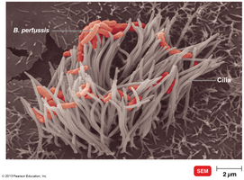
Pertussis is caused by Bordetella pertussis, a Gram-negative coccobacillus. The disease is characterized by severe coughing fits and is most dangerous in infants.
Stages: Catarrhal (cold-like), paroxysmal (violent cough), convalescence (recovery)
Virulence factors: Capsule, tracheal cytotoxin, pertussis toxin
Diagnosis: Clinical signs, culture on Bordet-Gengou agar, direct fluorescent antibody test
Prevention: DTaP vaccine (acellular)
Tuberculosis
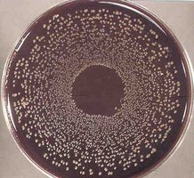
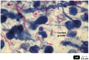
Tuberculosis (TB) is caused by Mycobacterium tuberculosis, an acid-fast rod. It is transmitted via airborne droplets and can cause chronic lung infection with systemic symptoms.
Symptoms: Weight loss, chronic cough, hemoptysis (coughing blood), fatigue
Diagnosis: Tuberculin skin test (Mantoux), chest X-ray, acid-fast staining, culture, NAATs
Treatment: Prolonged multi-drug therapy (isoniazid, rifampin, ethambutol, pyrazinamide)
Prevention: BCG vaccine (live attenuated M. bovis)


Pneumonias
Pneumonia is an infection of the lung parenchyma, caused by a variety of bacteria, viruses, and fungi. It is classified based on the causative agent and the anatomical region affected.
Bacterial causes: Streptococcus pneumoniae (most common), Haemophilus influenzae, Staphylococcus aureus, Legionella pneumophila, Mycoplasma pneumoniae
Symptoms: Fever, cough, chest pain, difficulty breathing, rust-colored sputum (pneumococcal)
Diagnosis: Chest X-ray, sputum culture, optochin-inhibition test, serology
Treatment: Depends on etiology (macrolides, cephalosporins, tetracyclines, erythromycin)
Prevention: Vaccines (pneumococcal, Hib)
Legionellosis
Legionellosis is caused by Legionella pneumophila, a Gram-negative rod found in water systems. It is transmitted by inhaling contaminated aerosols and can cause severe pneumonia, especially in older adults.
Symptoms: High fever, cough, pneumonia
Diagnosis: Culture on selective media, DNA probe
Treatment: Erythromycin
Psittacosis (Ornithosis)
Psittacosis is caused by Chlamydophila psittaci, an obligate intracellular Gram-negative bacterium. It is transmitted from birds to humans via inhalation of dried bird droppings.
Symptoms: Fever, headache, chills, sometimes delirium
Diagnosis: Growth in eggs or cell culture
Treatment: Tetracyclines
Chlamydial Pneumonia
Caused by Chlamydophila pneumoniae, this infection is transmitted human-to-human and is common in young people. It presents as a mild respiratory illness similar to mycoplasmal pneumonia.
Diagnosis: Serological tests
Treatment: Tetracyclines
Q Fever
Q fever is caused by Coxiella burnetii, with reservoirs in large mammals and transmission via ticks or unpasteurized milk. It presents as a mild respiratory disease but can have complications such as endocarditis.
Treatment: Doxycycline and chloroquine
Melioidosis
Melioidosis is caused by Burkholderia pseudomallei, found in soil in Southeast Asia and northern Australia. It can cause pneumonia, abscesses, and severe sepsis.
Treatment: Ceftazidime
Viral Diseases of the Respiratory System
COVID-19
COVID-19 is caused by SARS-CoV-2, a Betacoronavirus. It is transmitted via droplets and aerosols, with a wide range of symptoms from mild to severe, including fever, cough, and loss of taste or smell.
Diagnosis: Rapid antigen tests (ELISA), PCR
Treatment: Remdesivir, Paxlovid, molnupiravir, convalescent plasma
Prevention: mRNA, viral vector, and subunit vaccines
Influenza
Influenza is caused by Orthomyxoviruses, which have segmented RNA genomes and surface antigens hemagglutinin (HA) and neuraminidase (NA). Antigenic drift and shift contribute to seasonal epidemics and pandemics.
Symptoms: Chills, fever, headache, muscle aches
Treatment: Zanamivir, oseltamivir
Prevention: Annual multivalent vaccine
Respiratory Syncytial Virus (RSV)
RSV is a major cause of pneumonia in infants, leading to cell fusion (syncytium) in culture. It is diagnosed by serological tests and treated with ribavirin or palivizumab.
Fungal Infections of the Respiratory System
Histoplasmosis
Histoplasmosis is caused by Histoplasma capsulatum, a dimorphic fungus. Infection occurs via inhalation of conidia from soil, especially with bat droppings. It resembles miliary tuberculosis and is usually mild.
Diagnosis: Microscopy (yeast phase at 37°C), serology
Treatment: Amphotericin B
Coccidioidomycosis (Valley Fever)
Coccidioidomycosis is caused by Coccidioides immitis, found in desert soils of the American Southwest. It presents with chest pain, fever, and weight loss, and is diagnosed by serological tests.
Treatment: Amphotericin B
Pneumocystis Pneumonia
Pneumocystis pneumonia is caused by Pneumocystis jirovecii, an opportunistic pathogen affecting immunocompromised patients. It is diagnosed by microscopy and treated with trimethoprim.
Blastomycosis
Blastomycosis is caused by Blastomyces dermatitidis, found in soil in the Mississippi valley. It can cause abscesses, cutaneous ulcers, and extensive tissue damage.
Treatment: Amphotericin B
Summary Table: Major Bacterial Causes of Pneumonia
Pathogen | Gram Reaction | Key Features | Treatment |
|---|---|---|---|
Streptococcus pneumoniae | Gram-positive diplococcus | Encapsulated, rust-colored sputum | Macrolides, fluoroquinolones |
Haemophilus influenzae | Gram-negative coccobacillus | Requires X and V factors | Cephalosporins |
Mycoplasma pneumoniae | No cell wall | "Walking" pneumonia, PCR/serology | Tetracyclines |
Legionella pneumophila | Gram-negative rod | Waterborne, not person-to-person | Erythromycin |
Chlamydophila pneumoniae | Obligate intracellular | Mild, common in young people | Tetracyclines |
