 Back
BackMicrobial Diseases of the Respiratory System: Structure, Function, and Pathogenesis
Study Guide - Smart Notes
 Tailored notes based on your materials, expanded with key definitions, examples, and context.
Microbial Diseases of the Respiratory System
Overview of the Respiratory System
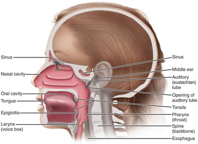
The respiratory system is divided into the upper and lower respiratory tracts, each with distinct anatomical structures and defense mechanisms against microbial invasion.
Upper respiratory system: Includes the nose, pharynx, middle ear, and eustachian tubes. Saliva and tears help protect mucosal surfaces.
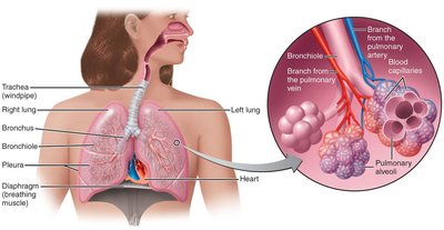
Lower respiratory system: Comprises the larynx, trachea, bronchial tubes, and alveoli. The ciliary escalator moves particles toward the throat, alveolar macrophages destroy microorganisms, and respiratory mucus protects mucosal surfaces.
Normal Microbiota of the Respiratory System
The upper respiratory tract harbors a diverse microbiota, including potentially pathogenic microorganisms. These normal microbiota suppress pathogens by competing for nutrients and producing inhibitory substances. The lower respiratory system is nearly sterile due to the effectiveness of the ciliary escalator and immune defenses.
Microbial Diseases of the Upper Respiratory System
Common Diseases and Their Characteristics
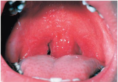
Pharyngitis: Inflammation of the throat, commonly known as a sore throat.
Laryngitis: Inflammation of the larynx, often resulting in voice loss.
Tonsillitis: Inflammation of the tonsils.
Sinusitis: Inflammation of the sinuses, often associated with headaches.
Epiglottitis: The most life-threatening disease of the upper respiratory system, often caused by Haemophilus influenzae type b.
Bacterial Diseases of the Upper Respiratory System
Streptococcal pharyngitis (strep throat): Caused by group A streptococci (Streptococcus pyogenes), which are resistant to phagocytosis and produce streptokinases and streptolysins. Symptoms include local inflammation, fever, tonsillitis, and enlarged lymph nodes. Diagnosis is by throat culture or rapid antigen detection tests.
Scarlet fever: Caused by erythrogenic toxin-producing strains of S. pyogenes. Characterized by a red rash and peeling skin.
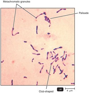
Diphtheria: Caused by Corynebacterium diphtheriae, a Gram-positive rod. It forms a tough grayish membrane in the throat, which can block air passage. The exotoxin can damage the heart and kidneys. Treated with antibiotics and antitoxin; prevented by the DTaP vaccine.
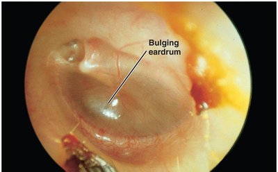
Otitis media: Infection of the middle ear, common in children. Caused by Streptococcus pneumoniae, nonencapsulated H. influenzae, Moraxella catarrhalis, S. pyogenes, and respiratory syncytial viruses. Treated with broad-spectrum penicillins.
Viral Diseases of the Upper Respiratory System
The Common Cold: Caused by over 200 different viruses, including rhinoviruses (30–50%), betacoronaviruses (10–15%), mastadenovirus, and enterovirus D68. Symptoms include sneezing, nasal secretion, and congestion. Antibiotics are ineffective; treatment is supportive.
Microbial Diseases of the Lower Respiratory System
Overview
Diseases of the lower respiratory system include bronchitis, bronchiolitis, and pneumonia. Pulmonary alveoli are often involved in pneumonia.
Bacterial Diseases of the Lower Respiratory System
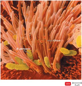
Pertussis (Whooping Cough): Caused by Bordetella pertussis, a Gram-negative coccobacillus. The bacterium produces a capsule for attachment to ciliated cells, destroys these cells, and shuts down the ciliary escalator. The disease progresses through three stages: catarrhal (cold-like), paroxysmal (violent coughing), and convalescence. Prevented by DTaP vaccine; treated with erythromycin or macrolides.
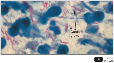
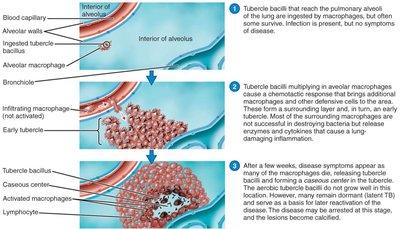
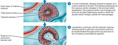
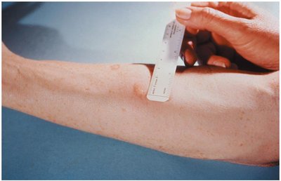
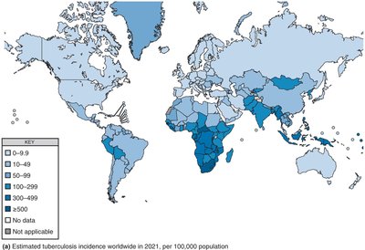
Tuberculosis: Caused by Mycobacterium tuberculosis, an acid-fast rod with a slow generation time. The bacterium is resistant to drying and antimicrobials due to mycolic acids in its cell wall. Pathogenesis involves formation of tubercles in the lungs, which may become calcified (Ghon’s complexes). If the immune response fails, the tubercle ruptures, releasing bacteria. Diagnosis includes the tuberculin skin test, x-ray, acid-fast staining, and PCR. Treatment requires prolonged multi-drug therapy due to slow growth and dormancy. MDR and XDR strains are a concern. The BCG vaccine is used in many countries but not widely in the U.S.

Bacterial Pneumonias
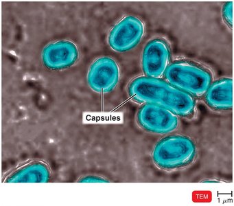
Pneumococcal pneumonia: Caused by Streptococcus pneumoniae, a Gram-positive encapsulated diplococcus. Symptoms include high fever, chest pain, and difficulty breathing. Diagnosis by optochin-inhibition test, bile solubility test, or capsular antigen in urine. Treated with macrolides; prevented by conjugated pneumococcal vaccine.
Haemophilus influenzae pneumonia: Caused by H. influenzae, a Gram-negative coccobacillus. Symptoms resemble pneumococcal pneumonia. Treated with third-generation cephalosporins; prevented by Hib vaccine.
Mycoplasmal pneumonia (walking pneumonia): Caused by Mycoplasma pneumoniae, which lacks a cell wall. Symptoms are mild but persistent. Diagnosis by PCR; treated with tetracyclines.
Legionellosis (Legionnaires’ disease): Caused by Legionella pneumophila. Grows in water systems and is transmitted by inhaling aerosols. Treated with azithromycin and macrolides.
Psittacosis (Ornithosis): Caused by Chlamydia psittaci, transmitted from birds. Treated with tetracyclines.
Chlamydial pneumonia: Caused by Chlamydia pneumoniae, transmitted person to person. Treated with azithromycin.
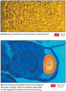
Q Fever: Caused by Coxiella burnetii, transmitted from animals or unpasteurized milk. Treated with doxycycline; chloroquine for chronic infections.
Melioidosis: Caused by Burkholderia pseudomallei, found in moist soils of Southeast Asia and northern Australia. Treated with ceftazidime.
Viral Diseases of the Lower Respiratory System
Major Viral Diseases
Viral pneumonia: Often a complication of influenza, measles, or chickenpox. Coronaviruses such as SARS-CoV, MERS-CoV, and SARS-CoV-2 (COVID-19) are notable causes.
COVID-19: Caused by SARS-CoV-2, a Betacoronavirus. Symptoms range from mild to severe, including fever, cough, and loss of smell/taste. Transmission is primarily airborne. Diagnosis by ELISA or PCR. Treated with antivirals such as remdesivir and Paxlovid. Vaccines include mRNA, virus-vector, and subunit types.
Respiratory Syncytial Virus (RSV): The most common viral respiratory disease in infants. Causes cell fusion (syncytium) in culture. Treated with palivizumab; vaccines are under development.
Influenza (Flu): Caused by Influenzavirus, which contains eight RNA segments and two types of spikes (HA and NA). Undergoes antigenic drift and shift, leading to epidemics and pandemics. Treated with neuraminidase inhibitors (zanamivir, oseltamivir); prevented by annual vaccination.
Fungal Diseases of the Lower Respiratory System
Major Fungal Diseases
Histoplasmosis: Caused by Histoplasma capsulatum, a dimorphic fungus. Acquired from airborne conidia in areas with bird or bat droppings. Treated with itraconazole.
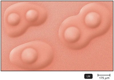
Coccidioidomycosis (Valley fever): Caused by Coccidioides immitis, found in desert soils. Forms spherules with endospores in tissues. Treated with fluconazole or itraconazole.
Pneumocystis pneumonia (PCP): Caused by Pneumocystis jirovecii, a yeastlike fungus. Primary indicator of AIDS. Treated with trimethoprim-sulfamethoxazole.
Blastomycosis: Caused by Blastomyces dermatitidis, a dimorphic fungus found in soil. Treated with itraconazole and amphotericin B.
Other fungi: Aspergillus fumigatus (aspergillosis), Rhizopus, and Mucor can cause respiratory disease, especially in immunocompromised individuals.
Summary Table: Selected Respiratory Diseases
Disease | Pathogen | Signs and Symptoms | Reservoir | Diagnosis | Treatment/Prevention |
|---|---|---|---|---|---|
Pneumococcal Pneumonia | Streptococcus pneumoniae | Infected alveoli fill with fluids; interferes with oxygen uptake | Humans | Optochin test, bile solubility, capsular antigen | Beta-lactam, macrolides; vaccine |
Haemophilus influenzae Pneumonia | Haemophilus influenzae | Resembles pneumococcal pneumonia | Humans | Isolation, special media | Cephalosporins; Hib vaccine |
Mycoplasmal Pneumonia | Mycoplasma pneumoniae | Mild, persistent respiratory symptoms | Humans | PCR | Tetracyclines |
Legionellosis | Legionella pneumophila | Potentially fatal pneumonia | Water | Culture on selective media | Azithromycin, macrolides |
Psittacosis | Chlamydia psittaci | Fever, headache, chills | Birds | Bacterial culture, PCR | Tetracyclines |
Chlamydial Pneumonia | Chlamydia pneumoniae | Mild respiratory illness | Humans | PCR | Azithromycin |
Q Fever | Coxiella burnetii | Mild respiratory disease; endocarditis | Large mammals, unpasteurized milk | Antibody titer | Doxycycline, chloroquine |
Melioidosis | Burkholderia pseudomallei | Pneumonia, abscesses, sepsis | Moist soil | Bacterial culture, PCR | Ceftazidime |
COVID-19 | SARS-CoV-2 | Fever, cough, loss of smell/taste | Humans | ELISA, PCR | Remdesivir, nirmatrelvir; vaccine |
RSV Disease | Human orthopneumovirus | Pneumonia in infants | Humans | Serological tests, PCR | Palivizumab |
Influenza | Influenzavirus | Chills, fever, headache, muscle aches | Humans, pigs, birds | Serological tests, PCR | Zanamivir, oseltamivir; vaccine |
Histoplasmosis | Histoplasma capsulatum | Resembles tuberculosis | Soil (Ohio/Mississippi River valleys) | Serological tests | Itraconazole |
Coccidioidomycosis | Coccidioides immitis | Fever, coughing, weight loss | Desert soils (U.S. Southwest) | Serological tests | Fluconazole, itraconazole |
Pneumocystis Pneumonia | Pneumocystis jirovecii | Pneumonia (immunocompromised) | Humans | Microscopy | Trimethoprim-sulfamethoxazole |
Blastomycosis | Blastomyces dermatitidis | Resembles bacterial pneumonia; tissue damage | Soil (Great Lakes/Mississippi River Valleys) | Isolation of pathogen | Itraconazole, amphotericin B |
Additional info: This summary integrates and expands upon the provided lecture slides and textbook images, ensuring a comprehensive, exam-ready overview of microbial diseases of the respiratory system for college-level microbiology students.
