 Back
BackMicrobial Diseases of the Respiratory System: Structure, Function, and Pathogenesis
Study Guide - Smart Notes
 Tailored notes based on your materials, expanded with key definitions, examples, and context.
Microbial Diseases of the Respiratory System
Structure and Function of the Respiratory System
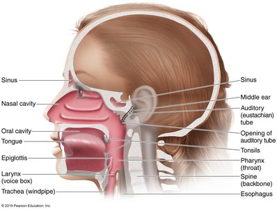
The respiratory system is divided into the upper and lower respiratory tracts, each with distinct anatomical features and protective mechanisms against microbial invasion.
Upper respiratory system: Includes the nose, pharynx, middle ear, and eustachian tube. Saliva and tears help protect mucosal surfaces from pathogens.
Lower respiratory system: Comprises the larynx, trachea, bronchial tubes, and alveoli. The ciliary escalator moves particles toward the throat, while alveolar macrophages destroy microorganisms in the lungs. Respiratory mucus also protects mucosal surfaces.
Normal Microbiota of the Respiratory System
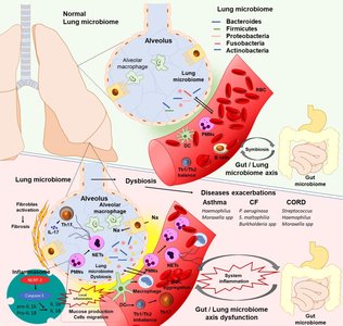
The normal microbiota of the respiratory tract plays a crucial role in suppressing pathogens by competing for nutrients and producing inhibitory substances. The lower respiratory system is nearly sterile due to these defenses.
Diseases of the Upper Respiratory System
Common Signs and Symptoms
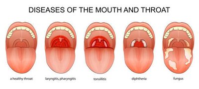
Pharyngitis: Sore throat and inflammation of the throat mucous membranes.
Laryngitis: Inflammation affecting the ability to speak.
Tonsilitis: Inflammation of the tonsils.
Sinusitis: Inflammation of the sinus mucous membranes, usually self-limiting.
Epiglottitis: The most life-threatening disease of the upper respiratory system.
Bacterial Infections
Strep Throat (Streptococcal Pharyngitis)
Etiology: Streptococcus pyogenes (Group A strep, GAS)
Virulence factors: Resistance to phagocytosis, streptokinases (lyse clots), streptolysins (cytotoxic).
Symptoms: Local inflammation, fever, tonsilitis, enlarged lymph nodes.
Diagnosis: Enzyme immunoassay (EIA).
Scarlet Fever
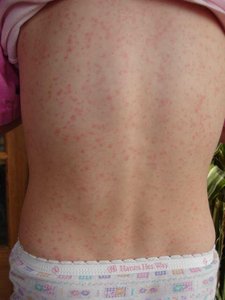
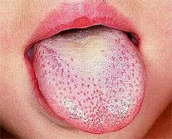
Etiology: Lysogenized S. pyogenes produces erythrogenic (reddening) toxin.
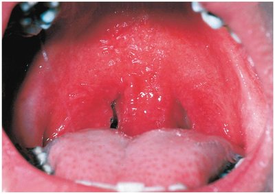
Symptoms: Red rash and 'strawberry tongue.'
Otitis Media (Middle Ear Infection)
Common pathogens: Streptococcus pneumoniae (35%), nonencapsulated Haemophilus influenzae (20-30%), Moraxella catarrhalis (10-15%), S. pyogenes (8-10%), Staphylococcus aureus (1-2%).
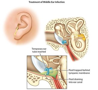
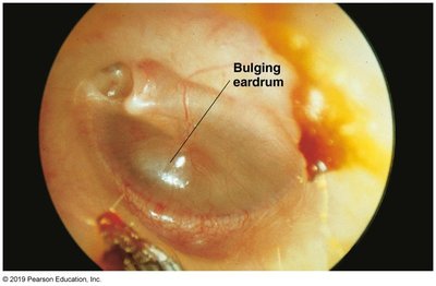
Pathogenesis: Pus formation increases pressure on the tympanic membrane; more common in children due to smaller eustachian tubes.
Treatment: Broad-spectrum antibiotics, ear tubes for drainage.
Viral Infections
Common Cold
Etiology: Rhinoviruses (30-50%), coronaviruses (10-15%).
Symptoms: Sneezing, nasal secretions, congestion, no fever.
Treatment: Cough suppressants and antihistamines; antibiotics are ineffective.
Complications: May progress to laryngitis and otitis media.
Diseases of the Lower Respiratory System
Common Signs and Symptoms
Bronchitis: Inflammation of large-medium sized airways (bronchi).
Bronchiolitis: Inflammation of small airways (bronchioles).
Pneumonia: Severe bronchitis involving pulmonary alveoli, leading to inflammation and impaired gas exchange.
Bacterial Infections
Pertussis (Whooping Cough)
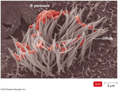
Etiology: Bordetella pertussis (Gram-negative coccobacillus, encapsulated).
Pathogenesis: Attaches to and destroys ciliated cells in the trachea, shutting down the ciliary escalator. Tracheal cytotoxin damages ciliated cells; pertussis toxin enters the bloodstream.
Stages:
Catarrhal stage: Similar to common cold.
Paroxysmal stage: Violent coughing, gasping for air.
Convalescence stage: Recovery, may last months; risk of brain damage in infants.
Prevention: DTaP vaccine (and Tdap booster).
Treatment: Erythromycin.
Recent trends: Cases rising due to breakdown in herd immunity, pathogen mutation, improved diagnostics, and lower long-term immunity from acellular vaccines.

Tuberculosis (TB)
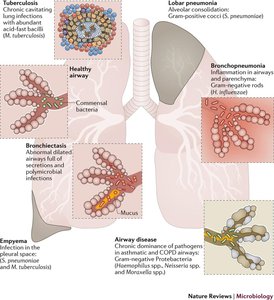
Etiology: Mycobacterium tuberculosis (acid-fast rod, obligate aerobe, slow-growing, lipid-rich cell wall).
Global impact: 9 million cases and 2 million deaths annually; 1/3 of the world's population infected.
Other species: Mycobacterium avium-intracellulare (HIV patients), Mycobacterium bovis (bovine TB, rare in US).
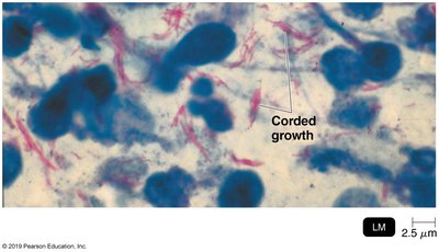
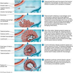
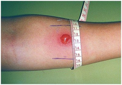
Diagnosis: Tuberculin skin test (delayed hypersensitivity), followed by X-ray, CT, acid-fast staining, culture, or rapid blood tests (IFN-γ, PCR).
Treatment: BCG vaccine (live, avirulent M. bovis, not widely used in US), prolonged multidrug chemotherapy (minimum 6 months).
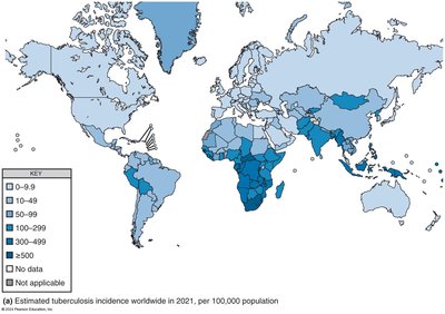
Pneumonia
Pneumonia is an infection of the lung parenchyma, with various etiologies and clinical presentations.
Types: Typical (e.g., Streptococcus pneumoniae), atypical (e.g., Mycoplasma pneumoniae, Legionella pneumophila), viral, and fungal.
Symptoms: Inflammation, difficulty breathing, dry cough, fever, chest pain.
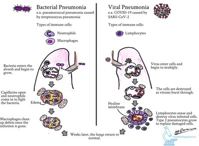
Bacterial Pneumonia
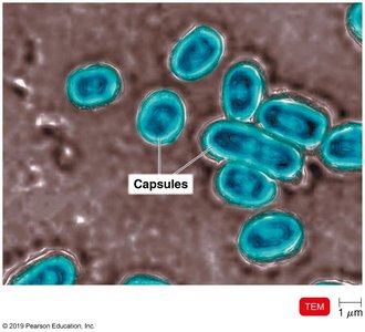
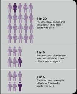
Typical (Pneumococcal) Pneumonia: Streptococcus pneumoniae (Gram-positive, encapsulated diplococci, 90 serotypes). Infects alveoli, causing fluid and RBC accumulation, interfering with oxygen uptake. Treated with macrolides or fluoroquinolones; prevented by conjugated vaccine.
Atypical Pneumonia: Haemophilus influenzae (Gram-negative coccobacillus), Mycoplasma pneumoniae (no cell wall, 'fried-egg' colonies), Legionella pneumophila (waterborne, not person-to-person). Symptoms are generally milder and may require special media for diagnosis. Treated with cephalosporins, tetracyclines, or macrolides as appropriate.
Viral Pneumonia
Common viruses: Influenza, measles, chickenpox, SARS-CoV-2 (COVID-19).
Pathogenesis: Often occurs as a complication of viral infection, leading to alveolar damage and impaired gas exchange.
Severe Acute Respiratory Syndrome (SARS) and COVID-19
Etiology: Coronaviruses (SARS-CoV, MERS-CoV, SARS-CoV-2).
Pathogenesis: Infects cells with ACE2 receptor (lungs, kidney, heart, etc.), causes endothelial disruption, coagulopathy, and cytokine storm, leading to pneumonia and multi-organ dysfunction.
Treatment: Palliative, monoclonal antibodies; prevention via mRNA or adenovirus-based vaccines.
Transmission: Droplets; highly contagious.
Respiratory Syncytial Virus (RSV)
Most common viral respiratory disease in infants. Nearly all children infected by age 2; causes cell fusion (syncytium), coughing, and wheezing. Diagnosed by serology; treated with ribavirin or palivizumab.
Influenza (Flu)
Etiology: Influenzavirus (RNA virus, 8 segments, lipid envelope).
Structure: Hemagglutinin (HA) spikes (attachment), neuraminidase (NA) spikes (release from cells).
Antigenic drift: Minor changes in HA/NA, eluding immunity.
Antigenic shift: Major changes, reassortment of RNA, leads to pandemics.
Prevention: Annual multivalent vaccine; treatment with neuraminidase inhibitors (zanamivir, oseltamivir).
Hantavirus
Forms: Hemorrhagic fever with renal syndrome (HFRS), hemorrhagic pulmonary syndrome (HPS).
Transmission: Aerosols from rodent urine/feces; HPS has high mortality (~40%).
Fungal Pneumonias
Pneumocystis Pneumonia
Etiology: Pneumocystis jirovecii; asymptomatic in immunocompetent, causes pneumonia in immunocompromised (AIDS indicator).
Pathology: Forms cysts in alveoli, releasing trophozoites.
Treatment: Trimethoprim-sulfamethoxazole.
Histoplasmosis
Etiology: Histoplasma capsulatum (dimorphic fungus).
Transmission: Inhalation of airborne conidia from bird/bat droppings; limited US range.
Treatment: Amphotericin B, itraconazole.
Coccidioidomycosis (Valley Fever)
Etiology: Coccidioides immitis (dimorphic fungus).
Transmission: Arthroconidia in desert soils; forms spherules with endospores in tissues.
Symptoms: Fever, cough, weight loss; <1% resemble TB.
Treatment: Amphotericin B, imidazole.
