 Back
BackMicrobial Diseases of the Skin and Eyes: Structure, Pathogenesis, and Clinical Manifestations
Study Guide - Smart Notes
 Tailored notes based on your materials, expanded with key definitions, examples, and context.
Microbial Diseases of the Skin and Eyes
Structure and Function of the Skin
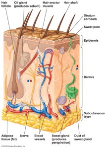
The skin is the largest organ of the human body and serves as a primary barrier against microbial invasion. Its structure and secretions play crucial roles in innate immunity and maintaining a balanced microflora.
Epidermis: The thin, outermost layer composed of epithelial cells, including the stratum corneum, which is rich in keratin and provides a tough, water-resistant barrier.
Dermis: The thicker, inner layer containing hair follicles, sweat glands, oil (sebaceous) glands, nerves, and blood vessels.
Secretions: Perspiration (contains lysozyme and antimicrobial peptides) and sebum (contains lipids, proteins, and fatty acids) inhibit microbial growth.
Normal Microbiota of the Skin
The skin's normal microbiota consists mainly of Gram-positive bacteria adapted to resist drying and high salt concentrations. While generally harmless, some can become opportunistic pathogens under certain conditions.
Common genera: Staphylococcus, Micrococcus, Propionibacterium acnes, Corynebacterium xerosis
These organisms can cause disease if the skin barrier is breached or the immune system is compromised.
Types of Skin Lesions
Skin lesions are not always indicative of infection but are important diagnostic features in microbial diseases.
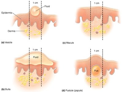
Vesicle: Small, fluid-filled lesion.
Bullae: Large (>1 cm) fluid-filled lesion.
Macule: Flat, reddened lesion.
Papule: Raised lesion.
Pustule: Raised lesion containing pus.
Bacterial Diseases of the Skin
Staphylococcal Skin Infections
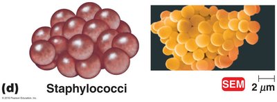
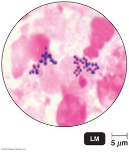
Staphylococcus species are Gram-positive cocci that form irregular clusters. They are classified based on coagulase production.
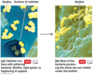
Coagulase-negative staphylococci: Staphylococcus epidermidis is common on skin and can form biofilms on medical devices, causing opportunistic infections.
Coagulase-positive staphylococci: Staphylococcus aureus is the most pathogenic, producing toxins that kill phagocytes and evade host defenses. It is notable for antibiotic resistance (MRSA, VRSA).
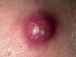
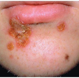
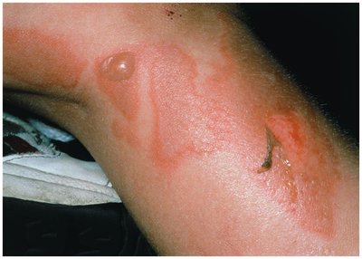
Diseases: Folliculitis (hair follicle infection), sty (eyelash), furuncle (boil), carbuncle (deep tissue infection), impetigo (highly contagious pustules), scalded skin syndrome (toxin-mediated separation of skin layers).
Streptococcal Skin Infections
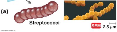
Streptococcus species are Gram-positive cocci that form chains and secrete various enzymes and toxins, including hemolysins.
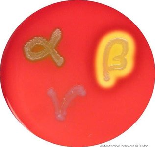
Beta-hemolytic streptococci: Streptococcus pyogenes (Group A) produces hemolysin, streptolysin, streptokinase, and DNAse.
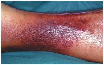
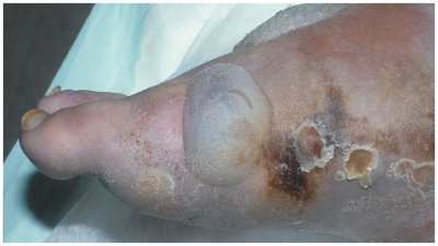
Diseases: Erysipelas (dermal infection, often after strep throat), necrotizing fasciitis ("flesh-eating" disease), and streptococcal toxic shock syndrome.
Pathogenesis: Exotoxin A acts as a superantigen, causing rapid tissue destruction and systemic shock.
Infections by Pseudomonads
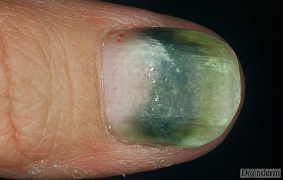
Pseudomonas species are Gram-negative rods known for their resistance to antibiotics and disinfectants. They produce exotoxins, endotoxins, and pigments such as pyocyanin.
Diseases: Dermatitis (self-limiting rash), otitis externa (swimmer’s ear), infections in burn victims, and biofilm-associated infections in cystic fibrosis patients and on catheters.
Virulence: Pyocyanin pigment generates reactive oxygen species and imparts a blue-green color to infected tissues.
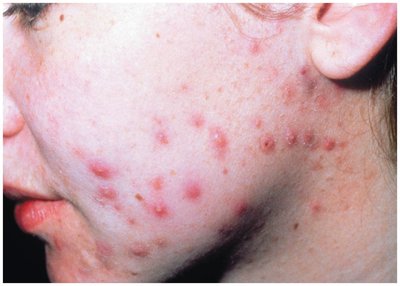
Acne
Acne is a common skin condition resulting from clogged hair follicles and sebaceous glands, often exacerbated by hormonal changes.
Etiology: Propionibacterium acnes, an anaerobic Gram-positive rod, metabolizes sebum and produces fatty acids that trigger inflammation.
Treatment: Antibiotics and topical agents to reduce sebum production.
Viral Diseases of the Skin
Chickenpox and Shingles
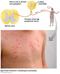
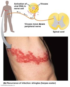
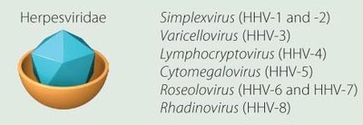
Both diseases are caused by the Herpesvirus varicella-zoster (HHV-3). Chickenpox is the primary infection, while shingles is a reactivation of latent virus.
Chickenpox: Virus enters via the respiratory tract, spreads to the skin, causing vesicular lesions that scab over.
Shingles: Latent virus in dorsal root ganglia reactivates, causing painful vesicles, usually on the torso.
Herpes Simplex Virus (HSV-1)
HSV-1 is a polyhedral, enveloped virus causing cold sores and herpetic keratitis. It establishes latency in the trigeminal nerve ganglion and can reactivate due to stress, fever, or UV exposure.
Clinical features: Painful vesicular lesions, often recurring in the same location.
Treatment: Antivirals such as acyclovir, docosanol, and hydrocolloid patches.
Measles (Rubeola)
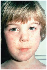
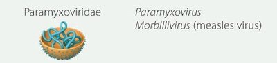
Measles is caused by the Morbillivirus (family Paramyxoviridae). It is a highly contagious disease with significant complications.
Symptoms: Fever, macular rash, cough, runny nose, sore throat.
Complications: Immune suppression, secondary infections, subacute sclerosing panencephalitis, blindness, deafness.
Prevention: MMR (measles, mumps, rubella) live-attenuated vaccine; herd immunity threshold is 95%.
Fungal Diseases of the Skin
Cutaneous Mycoses
Cutaneous mycoses are fungal infections limited to the hair, nails, or outer epidermis, caused by dermatophytes that digest keratin.
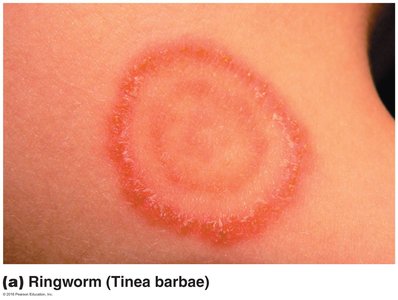
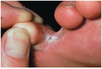
Tineas (Ringworm): Named by location: tinea cruris (groin, "jock itch"), tinea pedis (feet, "athlete's foot"), tinea unguium (nails).
Parasitic Diseases of the Skin
Scabies
Scabies is caused by the mite Sarcoptes scabiei, which burrows under the skin to lay eggs, causing intense itching and a variety of lesions. Secondary bacterial infections are common.
Diagnosis: Microscopic observation of mites, eggs, or fecal pellets.
Treatment: Topical scabicides.
Lice (Pediculosis)
Head lice (Pediculus humanus capitis) are adapted to grasp hair and feed on blood, causing itching due to sensitization to louse saliva.
Treatment: Topical insecticides and physical removal (combing).
Microbial Diseases of the Eye
Conjunctivitis
Conjunctivitis, or "pink eye," is inflammation of the conjunctiva caused by bacteria (e.g., Haemophilus influenzae), viruses (adenoviruses), or allergies.
Bacterial Diseases of the Eye
Ophthalmia neonatorum: Caused by Neisseria gonorrhoeae, acquired during childbirth, leading to pus formation and potential corneal ulceration. Treated with antibiotics.
Trachoma: Caused by Chlamydia trachomatis (non-STI strains), leading to conjunctivitis and trichiasis (inward turning of eyelashes). Treated with antibiotics or surgery.
Fungal Diseases of the Eye
Fungal keratitis is inflammation of the cornea, often following trauma, and can lead to blindness. Fusarium spp. are common causes, forming drug-resistant biofilms. The only approved treatment is natamycin.
Outbreaks: Contaminated contact lens solutions have caused outbreaks due to ineffective fungicides.
Summary Table: Major Microbial Diseases of the Skin and Eyes
Pathogen | Disease | Main Features | Treatment |
|---|---|---|---|
Staphylococcus aureus | Impetigo, boils, scalded skin syndrome | Pustules, bullae, exfoliative toxins | Antibiotics (resistance common) |
Streptococcus pyogenes | Erysipelas, necrotizing fasciitis | Beta-hemolysis, rapid tissue destruction | Beta-lactam antibiotics |
Pseudomonas aeruginosa | Dermatitis, otitis externa | Blue-green pigment, biofilms | Resistant to many drugs |
Propionibacterium acnes | Acne | Inflammatory papules, pustules | Antibiotics, topical agents |
Herpesvirus varicella-zoster | Chickenpox, shingles | Vesicular rash, latency in nerves | Supportive, antivirals for shingles |
HSV-1 | Cold sores | Recurrent vesicles, latency | Acyclovir, docosanol |
Morbillivirus | Measles | Macular rash, immune suppression | Supportive, MMR vaccine |
Dermatophytes | Tineas (ringworm) | Keratin digestion, ring-shaped lesions | Topical antifungals |
Sarcoptes scabiei | Scabies | Burrows, itching | Topical scabicides |
Pediculus humanus capitis | Head lice | Itching, visible nits | Topical insecticides, combing |
Neisseria gonorrhoeae | Ophthalmia neonatorum | Pus, corneal ulceration | Antibiotics |
Chlamydia trachomatis | Trachoma | Conjunctivitis, trichiasis | Antibiotics, surgery |
Fusarium spp. | Fungal keratitis | Corneal inflammation, biofilms | Natamycin |
