 Back
BackMicrobial Diseases of the Skin and Wounds: Structure, Microbiome, and Pathogenesis
Study Guide - Smart Notes
 Tailored notes based on your materials, expanded with key definitions, examples, and context.
Microbial Diseases of the Skin and Wounds
Structure and Function of the Skin
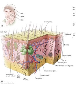
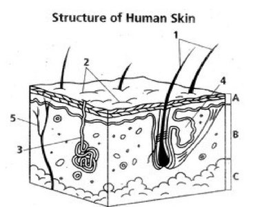
The skin is the largest organ of the human body and serves as a critical barrier against microbial invasion. It is composed of two main layers: the epidermis and the dermis, with a subcutaneous (hypodermis) layer beneath. The skin performs several essential functions that contribute to overall health and immunity.
Prevents excessive water loss
Regulates temperature
Assists in vitamin D formation
Involved in sensory phenomena
Acts as a barrier against microbial invaders
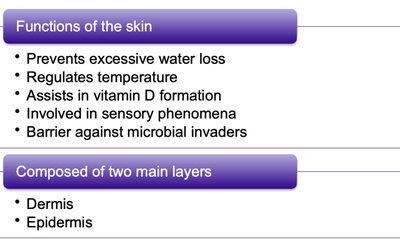
The epidermis is the outermost layer, primarily composed of keratinized cells, while the dermis contains connective tissue, blood vessels, nerves, and various glands. The hypodermis consists mainly of fat and connective tissue, providing insulation and cushioning.
Wounds and Their Role in Infection
Wounds are any trauma to the body's tissues, including cuts, scrapes, burns, bites, or surgical incisions. These injuries can disrupt the skin's barrier, allowing microbes to penetrate deeper tissues. While the immune system often eliminates these invaders, some infections can become severe or even fatal if not properly managed.
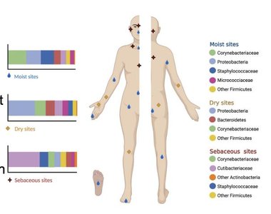
Microbiome of the Skin
The skin microbiome consists of a diverse community of microorganisms, including bacteria, fungi, and viruses. Most microbes do not survive long on the skin due to its high salt concentration, sebum (oil), and slightly acidic pH. However, certain species are adapted to thrive in this environment and play a protective role by competing with pathogens for nutrients and space, and by producing antimicrobial substances.
Common skin microbiota:
Yeasts: Malassezia
Gram-positive bacteria: Staphylococcus epidermidis, Micrococcus, Cutibacterium acnes
Diphtheroids: e.g., Cutibacterium acnes
Pathogenic microbes may cause disease if they penetrate the epidermis or if the immune system is compromised.

Bacterial Diseases of the Skin
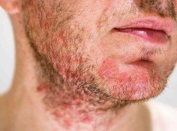
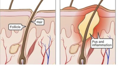
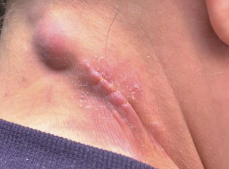
Folliculitis
Folliculitis is an infection of the hair follicle, often presenting as a pimple or, when occurring at the eyelid base, as a sty. If the infection spreads, it can form a furuncle (boil) or, when multiple furuncles coalesce, a carbuncle.
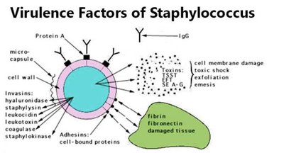
Pathogen: Most commonly caused by Staphylococcus species, especially S. aureus and S. epidermidis.
Virulence factors: Tolerance to salt and desiccation, production of enzymes and toxins.
Transmission: Direct contact or via fomites.
Diagnosis: Isolation of Gram-positive cocci in clusters from pus.
Treatment: Mupirocin or dicloxacillin; vancomycin for resistant strains (MRSA).
Prevention: Hand hygiene and proper hospital procedures.
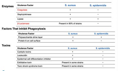
Comparison of Virulence Factors: S. aureus vs. S. epidermidis
Virulence Factor | S. aureus | S. epidermidis |
|---|---|---|
Coagulase | + | - |
Staphylokinase | + | - |
Lipase | + | + |
β-Lactamase | Present in 90% of strains | - |
Polysaccharide slime layer | + | + |
Protein A on cell surface | + | - |
Cytolytic toxins | + | - |
Leukocidin | + | - |
Epidermal cell differentiation inhibitor | Present in some strains | - |
Exfoliative toxin | Present in some strains | - |
Toxic shock syndrome toxin | Present in some strains | - |
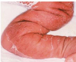
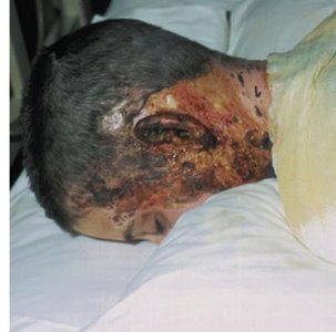
Staphylococcal Scalded Skin Syndrome (SSSS)
SSSS is caused by certain strains of S. aureus that produce exfoliative toxins. It is characterized by red, wrinkled skin that forms blisters and peels off in sheets, primarily affecting infants and young children. The disease is diagnosed by the characteristic sloughing of skin and treated with penicillin-derived drugs.
Impetigo, Erysipelas, and Cellulitis
These are common bacterial infections of the skin, often seen in children and individuals with compromised skin barriers.
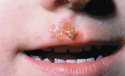
Impetigo: Red patches that develop into pus-filled vesicles, common in children. Caused by S. aureus (80%) and Streptococcus pyogenes (20%).
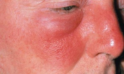
Erysipelas: Infection spreads to lymph nodes, causing reddening of the face, arms, or legs. Most often caused by Streptococcus.
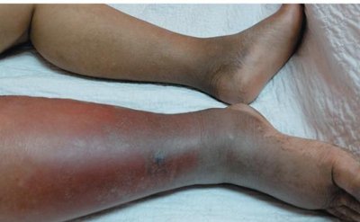
Cellulitis: Infection of the deeper dermis and subcutaneous fat, appearing as a red, swollen, hot area. Caused by Group A β-hemolytic streptococcus, Streptococcus pneumoniae, or S. aureus.
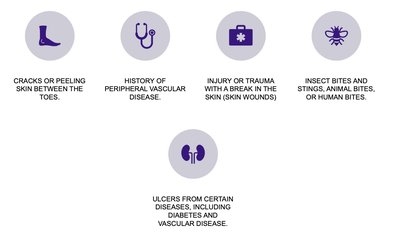
Risk Factors for Skin Infections
Cracks or peeling skin between the toes
History of peripheral vascular disease
Injury or trauma with a break in the skin
Insect bites and stings, animal bites, or human bites
Ulcers from diseases such as diabetes and vascular disease
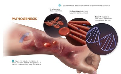
Necrotizing Fasciitis
Necrotizing fasciitis is a rapidly progressing infection caused primarily by Streptococcus pyogenes. It is characterized by redness, intense pain, swelling, fever, and tissue destruction. Early diagnosis and aggressive treatment, including surgical removal of affected tissue and broad-spectrum antimicrobials, are essential.
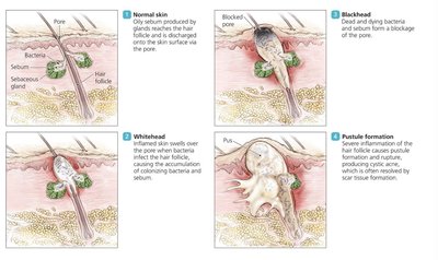
Acne
Acne is a common skin condition caused by Cutibacterium acnes (formerly Propionibacterium acnes), a Gram-positive, rod-shaped diphtheroid. It is associated with blocked hair follicles and inflammation, often beginning in adolescence.
Treatment: Antimicrobial drugs, exfoliating agents, Accutane for severe cases, and ultraviolet light therapy.
Pseudomonas Infection
Pseudomonas aeruginosa is a Gram-negative bacillus found in soil and moist environments. It is an opportunistic pathogen, particularly in burn victims, and produces a blue-green pigment (pyocyanin) in massive infections. Treatment is challenging due to multidrug resistance.
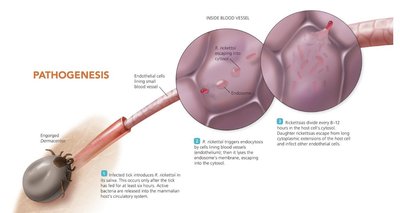
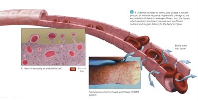
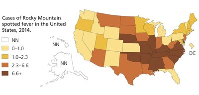
Spotted Fever Rickettsiosis (Rocky Mountain Spotted Fever)
Caused by Rickettsia rickettsii, this disease is transmitted by tick bites and is characterized by a non-itchy spotted rash (petechiae) and potential organ failure. Diagnosis is by serological testing, and prevention involves tick avoidance.
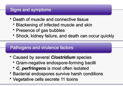
Gas Gangrene
Gas gangrene is caused by Clostridium perfringens and other Clostridium species. It is characterized by rapid tissue death, blackening of skin, gas bubble formation, and high mortality. Immediate surgical intervention and antimicrobials are required.
Viral Diseases of the Skin
Poxviruses
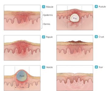
Poxviruses, including smallpox, orf, cowpox, and monkeypox, cause characteristic skin lesions that progress through several stages. Smallpox was eradicated through vaccination, but other poxviruses can still infect humans.
Herpes Infections
Herpes simplex viruses (HSV-1 and HSV-2) cause slow-spreading, painful, and itchy skin lesions. The viruses can remain latent and reactivate, causing recurrent outbreaks. Diagnosis is based on lesion appearance and immunoassays; treatment controls but does not cure the infection.
Warts
Warts are benign growths caused by human papillomaviruses (HPV). They can appear on various parts of the body and are transmitted by direct contact.
Chickenpox and Shingles
Chickenpox, caused by varicella-zoster virus (VZV), is highly contagious and characterized by widespread skin lesions. The virus can become latent and reactivate later in life as shingles, causing localized, painful rashes. Vaccines are available for both diseases.
Measles (Rubeola) and Rubella
Measles is marked by Koplik's spots and a widespread red rash, while rubella presents with a milder rash. Both are preventable by the MMR vaccine. Measles can lead to severe complications such as subacute sclerosing panencephalitis.
Parasitic Infestations of the Skin
Leishmaniasis
Leishmaniasis is caused by Leishmania species, transmitted by female sand flies. It presents as cutaneous, mucocutaneous, or visceral disease, with large painless skin lesions or involvement of mucous membranes and internal organs. Diagnosis is by microscopic identification, and prevention focuses on reducing exposure to sand flies.
Scabies
Scabies is caused by the mite Sarcoptes scabiei, leading to intense itching and rash, especially between the fingers and around the genitalia. Treatment involves mite-killing lotions and hygiene measures.
