 Back
BackChapter 19: Microbial Diseases of the Skin and Wounds
Study Guide - Smart Notes
 Tailored notes based on your materials, expanded with key definitions, examples, and context.
Microbial Diseases of the Skin and Wounds
Structure and Function of the Skin
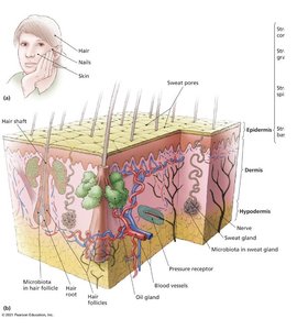
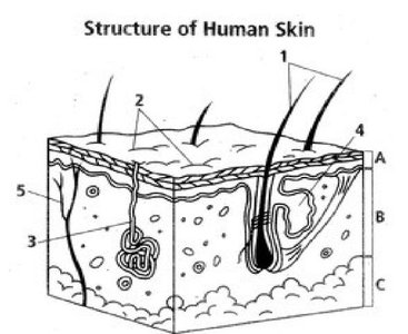
The skin is the largest organ of the human body and serves as a critical barrier against environmental hazards, including microbial invaders. It is composed of two main layers: the epidermis and the dermis, with a subcutaneous layer (hypodermis) beneath.
Prevents excessive water loss
Regulates temperature
Assists in vitamin D formation
Involved in sensory phenomena
Acts as a barrier against microbial invaders
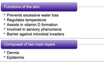
The skin consists of two main layers:
Dermis: Contains connective tissue, blood vessels, nerves, and glands.
Epidermis: Outermost layer, provides the primary barrier function.
Wounds and Microbial Entry
Wounds are any trauma to the body's tissues, such as cuts, scrapes, burns, bites, or surgical incisions. These breaches allow microbes to bypass the skin's barrier and potentially infect deeper tissues. While the immune system often eliminates these invaders, severe or fatal diseases can result if pathogens are not controlled.
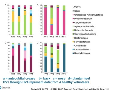
Microbiome of the Skin
The skin microbiome consists of a diverse community of microorganisms, including bacteria, fungi, and viruses. Most microbes do not survive long due to the skin's high salt concentration, sebum (oil), and slightly acidic pH. However, some harmless microbes persist and play protective roles.
Compete with pathogens for nutrients and space
Produce chemicals that inhibit other microbes
Cannot be completely removed by cleansing
Grow primarily in moist areas
Waste products can cause body odor
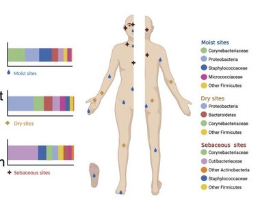
Common members of the skin microbiome include:
Yeast: Malassezia
Gram-positive bacteria: Staphylococcus epidermidis, Micrococcus, Cutibacterium acnes
Diphtheroids: e.g., Cutibacterium acnes
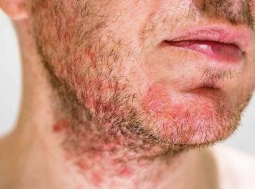
Bacterial Diseases of the Skin
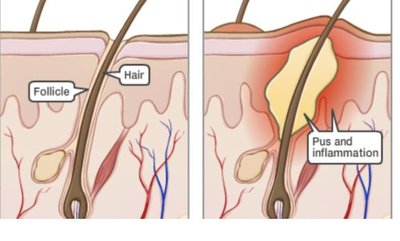
Folliculitis
Folliculitis is an infection of the hair follicle, often presenting as a pimple or, when at the eyelid base, a sty. If the infection spreads, it can form a furuncle (boil) or, when multiple furuncles coalesce, a carbuncle.
Pathogen: Most commonly caused by Staphylococcus species (Gram-positive cocci in clusters, facultatively anaerobic, salt and desiccation tolerant).
Common species: Staphylococcus epidermidis (rarely pathogenic), Staphylococcus aureus (transient colonizer, more virulent).
Transmission: Direct contact or fomites; can spread to blood and organs.
Diagnosis: Isolation of Gram-positive cocci in clusters from pus.
Treatment: Mupirocin or dicloxacillin; vancomycin for resistant strains (MRSA).
Prevention: Hand hygiene and proper hospital procedures.
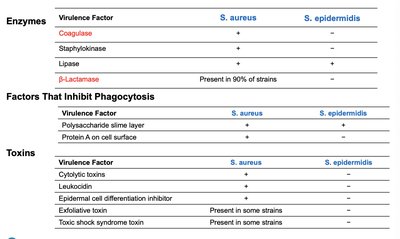
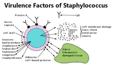
Comparison of Virulence Factors: S. aureus vs. S. epidermidis
Virulence Factor | S. aureus | S. epidermidis |
|---|---|---|
Coagulase | + | - |
Staphylokinase | + | - |
Lipase | + | + |
β-Lactamase | Present in 90% of strains | - |
Polysaccharide slime layer | + | + |
Protein A on cell surface | + | - |
Cytolytic toxins | + | - |
Leukocidin | + | - |
Epidermal cell differentiation inhibitor | Present in some strains | - |
Exfoliative toxin | Present in some strains | - |
Toxic shock syndrome toxin | Present in some strains | - |
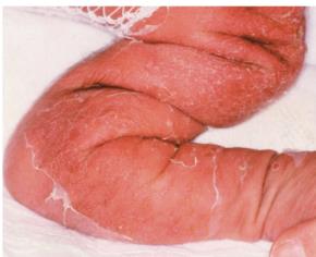
Staphylococcal Scalded Skin Syndrome (SSSS)
SSSS is caused by certain strains of S. aureus that produce exfoliative toxins. It is characterized by red, wrinkled skin that forms blisters and peels off in sheets. The dermis is unaffected, so no scarring occurs. Most common in infants and young children, it is transmitted person-to-person.
Treatment: Penicillin-derived drugs
Prevention: Difficult due to widespread presence of S. aureus
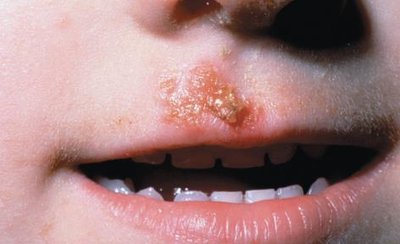
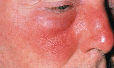
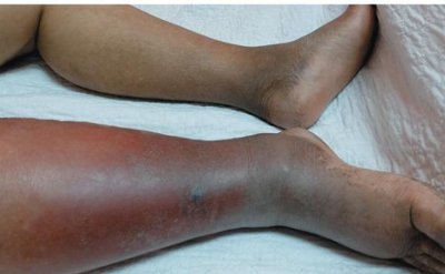
Impetigo, Erysipelas, and Cellulitis
These are common bacterial skin infections, especially in children and the elderly.
Impetigo: Red patches on face/limbs, develop into pus-filled vesicles. Caused by S. aureus (80%) and Streptococcus pyogenes (20%). Treated with oral/topical antimicrobials.
Erysipelas: Infection spreads to lymph nodes, causing reddening of face, arms, or legs. Most often caused by Streptococcus. Treated with penicillin.
Cellulitis: Infection of deeper dermis and subcutaneous fat, appears as a red, swollen, hot area. Caused by Group A β-hemolytic streptococcus, Streptococcus pneumoniae, or S. aureus. Treated with oral or IV antibiotics.
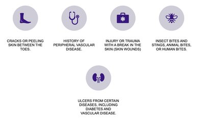
Risk Factors for Skin Infections
Cracks or peeling skin between toes
Peripheral vascular disease
Injury or trauma with skin break
Insect/animal/human bites
Ulcers from diseases like diabetes
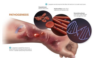
Necrotizing Fasciitis
Necrotizing fasciitis is a rapidly progressing infection of the fascia, often called "flesh-eating disease." Most cases are caused by S. pyogenes, which produces enzymes and toxins that destroy tissue.
Symptoms: Redness, intense pain, swelling, fever, nausea, malaise, mental confusion
Treatment: Early diagnosis, surgical removal of affected tissue, broad-spectrum antimicrobials
Prevention: Difficult due to common presence of S. pyogenes
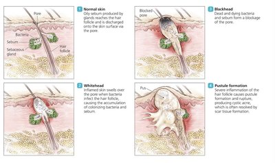
Acne
Acne is commonly caused by Cutibacterium acnes (formerly Propionibacterium acnes), a Gram-positive, rod-shaped diphtheroid. It is a normal member of the skin microbiome and typically begins in adolescence.
Treatment: Antimicrobial drugs, exfoliating agents, Accutane for severe cases, ultraviolet light
Pseudomonas Infection
Pseudomonas aeruginosa is a Gram-negative bacillus found in soil and moist environments. It rarely causes disease in healthy individuals but can infect burn victims, leading to fever, chills, shock, and blue-green pigment (pyocyanin) in massive infections.
Treatment: Combination antimicrobials; debridement of burns is essential
Prevention: Difficult due to widespread presence and multidrug resistance

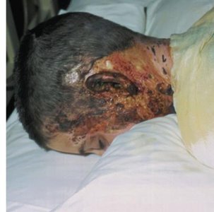
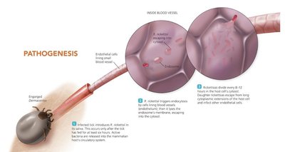
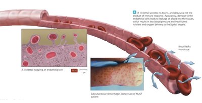
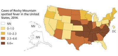
Spotted Fever Rickettsiosis (Rocky Mountain Spotted Fever)
Caused by Rickettsia rickettsii, a Gram-negative intracellular parasite, this disease is transmitted by tick bites. It presents with a non-itchy spotted rash (petechiae) and can lead to organ failure.
Treatment: Antimicrobials
Prevention: Tick repellents, avoiding tick-infested areas
Cutaneous Anthrax
Caused by Bacillus anthracis, cutaneous anthrax is characterized by a black, painless ulcer called an eschar. It is treated with antimicrobials and prevented by controlling the disease in animals.
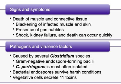
Gas Gangrene
Gas gangrene is caused by Clostridium species, especially C. perfringens. It involves death of muscle and connective tissue, blackening of skin, gas bubbles, and can lead to shock and death.
Treatment: Rapid surgical removal of dead tissue, antitoxin, antimicrobials
Prevention: Proper wound cleaning
Viral Diseases of the Skin and Wounds
Poxviruses
Poxviruses, including smallpox, orf, cowpox, and monkeypox, are DNA viruses that cause skin lesions progressing through several stages. Smallpox was eradicated globally, but other poxviruses still occur.
Transmission: Smallpox by inhalation; others by direct contact
Treatment: Immediate vaccination
Prevention: Vaccination (discontinued for smallpox in the 1970s)
Herpes Infections
Caused by human herpesviruses 1 and 2, these infections produce slow-spreading, painful, and itchy skin lesions. The viruses can remain latent and reactivate, causing recurrent lesions.
Treatment: Chemotherapeutic drugs (control, not cure)
Prevention: Gloves for healthcare workers
Warts
Warts are benign growths caused by papillomaviruses. They are transmitted by direct contact and can be removed by various methods, though recurrence is common.
Chickenpox and Shingles
Chickenpox is a highly contagious disease caused by varicella-zoster virus (VZV), characterized by lesions on the trunk and body. The virus can become latent and reactivate as shingles, causing localized painful lesions.
Treatment: Symptom relief, antiviral medications
Prevention: Vaccines for chickenpox and shingles
Rubella and Measles (Rubeola)
Rubella and measles are viral diseases causing characteristic rashes. Measles is marked by Koplik's spots and red lesions, with rare but severe complications. Both are preventable by vaccination (MMR vaccine).
Erythema Infectiosum (Fifth Disease) and Roseola
Erythema infectiosum causes a "slapped cheek" rash, while roseola is characterized by a rose-colored rash in children, caused by human herpesvirus 6 (HHV-6).
Parasitic Infestations of the Skin
Leishmaniasis
Leishmaniasis is caused by Leishmania species, protozoa transmitted by female sand flies. It presents as cutaneous, mucocutaneous, or visceral forms, with the latter being most severe.
Treatment: Antimicrobials for severe cases
Prevention: Reducing exposure to reservoir hosts
Scabies
Scabies is caused by the mite Sarcoptes scabiei, leading to intense itching and rash, especially between fingers, around genitalia, and on wrists, elbows, and knees. Treated with mite-killing lotions and hygiene measures.
