 Back
BackMicrobial Diseases of the Skin and Eyes: Structure, Defenses, and Pathogenesis
Study Guide - Smart Notes
 Tailored notes based on your materials, expanded with key definitions, examples, and context.
Microbial Diseases of the Skin and Eyes
Introduction
The skin and eyes serve as critical barriers against microbial invasion. However, various pathogens can overcome these defenses, leading to a range of diseases. This chapter explores the structure and defenses of the skin and mucous membranes, the normal microbiota, and the major bacterial diseases affecting these tissues.
Skin and Mucous Membrane Structure & Host Defenses
Skin Anatomy and Physiology
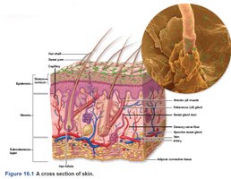
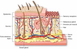
The skin is the largest organ of the body and acts as the first line of defense against pathogens. Its structure and secretions create an inhospitable environment for most microbes.
Epidermis: Outermost layer, replaced monthly, provides a physical barrier.
Dermis: Contains hair follicles, sweat, and oil glands, which can be entry points for microbes if breached.
Secretions: Sweat contains lysozyme (hydrolyzes peptidoglycan), antimicrobial fatty acids, defensins, and salt, all of which inhibit microbial growth.
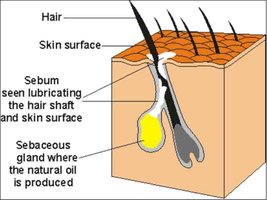
Sebum: Provides nutrients for some microbes but also contains antimicrobial properties.
Mucous Membranes (Mucosa)
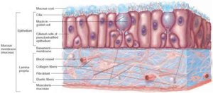
Mucous membranes line body cavities such as the respiratory, gastrointestinal, urinary, and reproductive tracts. They provide both physical and chemical barriers to infection.
Mucus: Traps particles, including microbes.
Cilia: Sweep debris and microbes out of the body.
Acidity: Many mucosal surfaces are acidic, limiting microbial populations.
Lysozyme: Present in tears and mucus, destroys cell walls of Gram-positive bacteria.
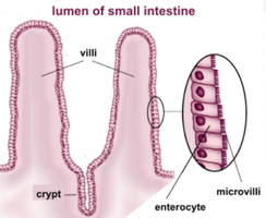
Microvilli: Increase surface area for absorption and defense.
Host Defenses Summary Table
Defenses | Normal Biota |
|---|---|
Keratizined surface, sloughing, low pH, high salt, lysozyme, antimicrobial peptides | Streptococcus, Staphylococcus, Corynebacterium, Propionibacterium, Pseudomonas, Lactobacillus, Candida |
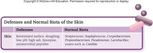
Normal Microbiota of the Skin
Composition and Ecological Roles
The skin's normal microbiota prevents colonization by pathogens and can become opportunistic pathogens if the immune system is compromised.
Mostly Gram-positive, salt-tolerant, dry-resistant bacteria: Staphylococci, Micrococci, Diphtheroids
Fungi: Malassezia species (yeasts), Candida albicans
Ecological roles: Compete with pathogens, produce antimicrobial substances, and modulate immune responses.
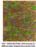
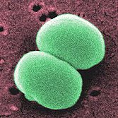
Key Members of Skin Microbiota
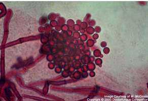
Diphtheroids: Pleomorphic, non-motile Gram-positive rods. Corynebacterium xerosis (aerobes on skin surface), Cutibacterium acnes (anaerobes in hair follicles, associated with acne and odor).
Staphylococci/Micrococci: Universally present, salt-tolerant. Staphylococcus epidermidis is the most common skin bacterium.
Fungi: Malassezia (dandruff), Candida albicans (opportunistic infections).
Types of Skin Lesions
Classification of Skin Rashes and Lesions
Skin lesions are classified based on their appearance and location. Understanding these types is essential for diagnosing skin diseases.
Exanthem: Rash on the skin (e.g., measles).
Enanthem: Rash on mucous membranes (e.g., inside the mouth).
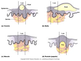
Vesicles: Small, fluid-filled lesions (<1 cm).
Bullae: Larger fluid-filled lesions (>1 cm).
Macules: Flat, red lesions.
Papules: Raised, solid lesions (no fluid).
Pustules: Raised lesions containing pus.
Bacterial Diseases of the Skin
Staphylococcal Skin Infections
Staphylococci are spherical, Gram-positive bacteria that often grow in clusters. They are major causes of skin infections, with Staphylococcus aureus and Staphylococcus epidermidis being the most significant species.
S. aureus: Coagulase-positive, pathogenic, produces penicillinase (antibiotic resistance), secretes leukocidin, resists lysozyme and opsonization, survives in phagolysosome, produces superantigens (TSST-1, enterotoxins).
S. epidermidis: Coagulase-negative, common skin microbiota, forms biofilms, opportunistic pathogen in healthcare settings.
Transmission: Often from nasal passages to skin, enters through hair follicles or skin breaks.
Types of Staphylococcal Skin Infections
Folliculitis: Infection of hair follicles.
Sty: Folliculitis of an eyelash.
Furuncle (Boil): Localized pus surrounded by inflamed tissue; can progress to carbuncle if not contained.
Carbuncle: Deep inflammation of tissue under the skin, may cause fever and illness.
Impetigo: Highly contagious, crusty sores, common in children, can be nonbullous (self-limiting) or bullous (caused by exfoliative toxin A, seen in newborns).
Scalded Skin Syndrome (SSSS): Caused by exfoliative toxin B, leads to widespread blistering and skin peeling, risk of secondary infections.
Toxic Shock Syndrome (TSS): Sudden fever, rash, hypotension, organ failure; caused by TSST-1 superantigen, associated with tampon use and wound infections.
Streptococcal Skin Infections
Streptococci are Gram-positive cocci that grow in chains and are classified by hemolytic enzymes and cell wall antigens. The most important group is Group A β-hemolytic streptococci (GAS), especially Streptococcus pyogenes.
Virulence factors: Hemolysins (lyse RBCs), streptokinase (dissolves clots), hyaluronidase (dissolves connective tissue), M protein (attachment, antiphagocytic).
Diseases: Localized skin infections, impetigo (sometimes co-infection with S. aureus), necrotizing fasciitis ("flesh-eating disease"), streptococcal toxic shock syndrome.
Necrotizing fasciitis: Rapid tissue destruction, high mortality, requires broad-spectrum antibiotics.
Infections by Pseudomonads
Pseudomonas aeruginosa is an aerobic, Gram-negative rod and a model opportunistic pathogen. It is notable for its resistance to antibiotics and its ability to form biofilms.
Pyocyanin: Exotoxin that produces blue-green pus.
Diseases: Pseudomonas dermatitis (swimming pools, hot tubs), otitis externa ("swimmer's ear"), respiratory infections, post-burn infections.
Treatment: Difficult due to resistance; requires sensitivity testing and high-dose IV antibiotics.
Acne
Acne is classified based on severity and underlying cause. It typically affects areas with dense sebaceous glands.
Comedonal (Mild) Acne: Blocked sebaceous ducts, forms whiteheads and blackheads. Treated with topical retinoids and salicylic acid.
Inflammatory (Moderate) Acne: Caused by Cutibacterium acnes, leads to pustules and papules. Treated with benzoyl peroxide, antibiotics, and light therapy.
Nodular Cystic (Severe) Acne: Deep, pus-filled nodules and cysts, can cause scarring. Treated with oral antibiotics and isotretinoin (Accutane®), which is teratogenic.
Review Questions and Key Concepts
What perspiration factors discourage microbial growth? High salt concentration, lysozyme, antimicrobial peptides, and fatty acids.
Are skin bacteria more likely to be Gram-positive or Gram-negative? Gram-positive (e.g., Staphylococcus, Streptococcus).
Which bacterial species features the virulence factor M protein? Streptococcus pyogenes.
What is the common name for otitis externa? Swimmer's ear.
