 Back
BackMicrobial Diseases of the Skin and Eyes: Structure, Pathogenesis, and Clinical Manifestations
Study Guide - Smart Notes
 Tailored notes based on your materials, expanded with key definitions, examples, and context.
Diseases of the Skin and Eyes
Introduction
The skin and eyes serve as important barriers against microbial invasion. However, various microorganisms can overcome these defenses, leading to a range of infectious diseases. This chapter explores the structure of the skin, its normal microbiota, and the major bacterial, viral, fungal, and arthropod-associated diseases affecting the skin and eyes.
Skin: Structure and Defense Mechanisms
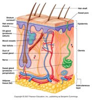
Properties of the Skin
Salty, dry, and low pH environment: These properties inhibit microbial growth.
Lysozymes: Enzymes that hydrolyze peptidoglycan, breaking down bacterial cell walls.
Normal microbiota: Includes Gram-positive, salt-tolerant bacteria such as Staphylococci, Micrococci, and diphtheroids.
Mucous Membranes
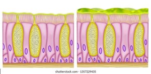
Mucus secretion: Traps foreign invaders.
Ciliary escalator: Some cells have cilia that move mucus and trapped particles out of the body.
Common Signs and Symptoms of Skin Diseases
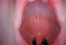
Exanthem and Enanthem
Exanthem: Skin rash arising from disease conditions.
Enanthem: Rash on mucous membranes (e.g., inside the mouth).
Lesions and Pus Formation
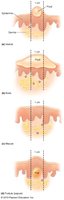
Lesions: Formed from fluid within skin layers; differences depend on the layer affected.
Pus: Protein-rich fluid containing dead leukocytes and cellular debris.
Types of Skin Lesions
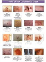
Lesions can be classified based on appearance and underlying pathology (e.g., macule, papule, vesicle, pustule, ulcer, etc.).
Bacterial Infections of the Skin
Staphylococcus Species
Staphylococcus epidermidis: Gram-positive, normal flora, opportunistic.
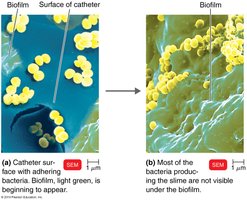
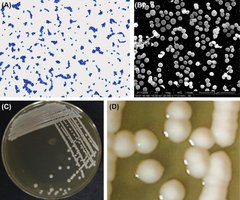
Staphylococcus aureus: Gram-positive, often found in nasal passages, most pathogenic of staphylococci, causes nosocomial infections, several antibiotic-resistant strains (e.g., MRSA).
Virulence factors: Coagulase (clots fibrin), toxins (leukocidins, hemolysins, exfoliative toxin, enterotoxins).
Common Staphylococcal Infections
Folliculitis: Infection of hair follicles.
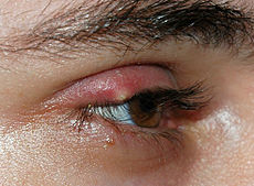
Sty (stye): Folliculitis of an eyelash.
Furuncle: Abscess/boil; localized pus surrounded by inflamed tissue.
Carbuncle: Inflammation of tissue under the skin, can lead to fever and generalized illness.
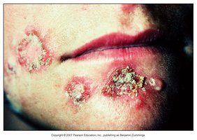
Impetigo: Highly contagious skin infection, primarily in children.
Scalded Skin Syndrome (SSS): Toxin-mediated loss of skin adhesion.
Toxic Shock Syndrome (TSS): TSST-1 superantigen triggers massive cytokine release, leading to shock and potentially death.
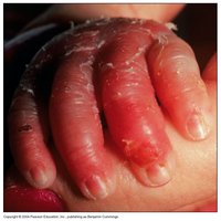
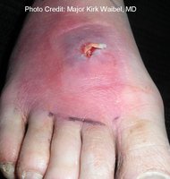
MRSA (Methicillin-resistant Staphylococcus aureus)
HA-MRSA: Hospital-acquired, serious for immunocompromised patients.
CA-MRSA: Community-acquired, more deadly, secretes high levels of modulins (peptides that destroy neutrophils).
Streptococcus Species
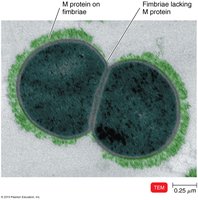
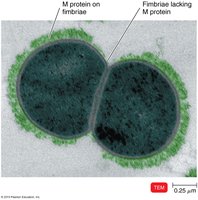
Streptococcus pyogenes: Gram-positive, grows in chains, catalase negative, Group A Streptococci (GAS), produces beta-hemolysins, M proteins on cell walls.
Virulence Factors
Hemolysins: Destroy red blood cells (alpha, beta, gamma types).
Leukocidins: Destroy leukocytes.
M-protein: Antiphagocytic, aids in adherence, antigenic variation, degrades complement protein C3b.
Streptokinases: Dissolve blood clots.
Streptococcal pyrogenic exotoxins (SPEs): Superantigens causing fever and hypersensitivity (e.g., scarlet fever, STSS).
Common Streptococcal Infections
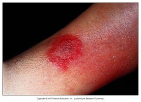
Erysipelas: Erythema affecting the dermal layer, sometimes includes lymphatics.
Impetigo: Also caused by Streptococcus.
Necrotizing Fasciitis
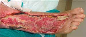
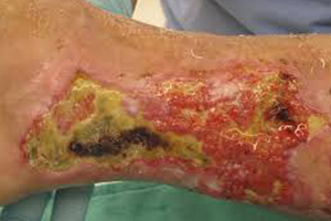
Caused by GAS producing streptokinases, hyaluronidase, and SPE superantigens.
Results in rapid death of soft tissue, can be fatal if untreated.
Pseudomonads
Pseudomonas aeruginosa: Gram-negative, aerobic rod, produces blue-green pus (pyocyanin).
Pseudomonas dermatitis: Associated with swimming pools and hot tubs.
Otitis externa: Swimmer’s ear infection.
Major concern for nosocomial infections, especially in burn patients.
Propionibacterium
Propionibacterium acnes: Gram-positive, anaerobic rod, causes inflammatory acne by metabolizing sebum.
Treatment: Antibiotics, benzoyl peroxide, visible (blue) light therapy.
Viral Infections of the Skin
Papillomaviruses (HPV)
Over 50 types; cause warts, transmitted by contact.
HPV-16 associated with cancers; vaccine reduces cervical cancer risk by 85–90%.
Epidermodysplasia verruciformis ("tree man" disease) is a rare susceptibility to HPV.
Poxviruses
Cause ulcers, macular rash, fever; transmitted via respiratory route.
Smallpox (variola virus): Variola major (~20% mortality), variola minor (<1% mortality).
First disease with a vaccine (Edward Jenner, 1796).
Monkeypox: Zoonotic, milder than smallpox, recent outbreaks.
Herpesviruses
Varicella-zoster virus (HHV-3): Causes chickenpox (varicella) and shingles (zoster); latent in dorsal root ganglion.
HSV-1: Oral herpes (cold sores, blisters).
HSV-2: Genital herpes.
Other forms: Herpes gladiatorum (skin), herpes whitlow (fingers), herpes encephalitis (brain, high fatality).
Treatment: Antivirals such as acyclovir.
Measles Virus (Rubeola)
Highly contagious; transmitted via respiratory route.
Symptoms: Koplik’s spots (enanthem in mouth), macular rash, fever.
Complications: Immune amnesia, encephalitis (1/1,000 cases).
Vaccine: MMR (measles, mumps, rubella).
German Measles Virus (Rubella)
Milder than measles; macular rash, fever.
Congenital rubella syndrome: Severe fetal damage (deafness, cataracts, learning disabilities).
Vaccine: MMR.
Parvoviruses and Roseola
Human parvovirus B19: Erythema infectiosum (fifth disease), flu-like symptoms, "slapped cheek" rash.
HHV-6 and HHV-7: Roseola (sixth disease), high fever, rash lasting 1–2 days.
Fungal Infections of the Skin
Cutaneous Mycoses
Fungi that metabolize keratin (dermatomycoses, tineas/ringworm).
Trichophyton: Infects hair, skin, nails.
Epidermophyton: Infects skin, nails.
Microsporum: Infects hair, skin.
Treatment: Oral griseofulvin, topical miconazole.
Subcutaneous Mycoses
Sporotrichosis (Sporothrix schenckii): Enters via puncture wound, causes nodular lesions, can spread if untreated. Treated with potassium iodide (KI).
Candidiasis
Candida albicans: Causes oral thrush (white patches, soreness), urogenital yeast infections (itching, discharge).
Predisposing factors: Antibiotic therapy, immunosuppression.
Treatment: Miconazole, nystatin, diflucan.
Arthropod Infections of the Skin
Scabies
Sarcoptes scabiei (mite): Burrows in skin to lay eggs, causes itching and rash.
Transmitted via contact and fomites (clothes, bedding).
Treatment: Topical insecticides.
Pediculosis (Lice)
Pediculus humanus capitis (head louse): Causes itching, mild reaction to bites, swelling of lymph nodes.
Transmitted via contact; females lay eggs (nits) on hair.
Treatment: Topical insecticides, removal of eggs.
Diseases of the Eyes
Bacterial Infections
Conjunctivitis (pinkeye): Most commonly caused by Haemophilus influenzae; symptoms include swelling of conjunctiva and red eyes. Transmitted via contact, associated with unsanitary conditions. Treated with topical antibiotics.
Ophthalmia neonatorum: Caused by Neisseria gonorrhoeae, transmitted to newborns during birth, can lead to blindness. Prevented with antibiotics or silver nitrate.
Inclusion conjunctivitis and trachoma: Caused by Chlamydia trachomatis, can lead to blindness, transmitted via birth canal, fomites, or arthropods. Treated with tetracycline.
Protozoan Infections
Acanthamoeba keratitis: Caused by Acanthamoeba spp., slowly destroys the cornea, leading to blindness. Transmitted via contaminated water or unsanitary contact lenses.
