 Back
BackMicrobial Diseases of the Skin and Wounds: Structure, Microbiome, and Pathogenesis
Study Guide - Smart Notes
 Tailored notes based on your materials, expanded with key definitions, examples, and context.
Microbial Diseases of the Skin and Wounds
Structure and Function of the Skin
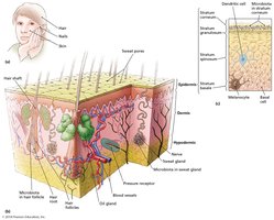
The skin is the largest organ of the human body and serves as a critical barrier against microbial invasion. It is composed of two main layers: the superficial epidermis and the deeper dermis. The skin prevents excessive water loss, regulates temperature, is involved in sensory phenomena, assists in vitamin D formation, and acts as a barrier to pathogens.
Epidermis: Outermost layer, composed of dead, keratinized cells, providing a tough barrier.
Dermis: Contains connective tissue, blood vessels, nerves, hair follicles, and glands.
Functions: Sensory detection, temperature regulation, and protection from mechanical and microbial threats.
Wounds and Their Impact on Skin Integrity
Wounds are trauma to any tissue of the body that breach the mechanical barrier provided by intact skin. Examples include cuts, scrapes, burns, bites, and surgical incisions. Wounds allow microbes to infect deeper tissues, but the body responds by forming blood clots and initiating tissue repair. Most wound infections are eliminated by immune defenses, but some can overwhelm the body and cause severe disease.
Why the Skin Surface Is Inhospitable to Most Microbes
The skin is covered with salt (from sweat) and sebum (an oily lipid), both of which have antimicrobial properties.
The outermost layer consists of dead, keratinized cells, which are regularly shed, removing attached microbes.
Antimicrobial chemicals in sweat and sebum inhibit microbial growth.
Microbiome of the Skin
Composition and Role of Skin Microbiota
The skin microbiome consists of bacteria and yeasts that tolerate the harsh conditions of the epidermis, hair follicles, and sweat ducts. These microbes are generally harmless and compete with pathogens for nutrients and space, producing chemicals that inhibit the growth of other microbes. Vigorous scrubbing reduces but does not eliminate these organisms, as they recolonize from deeper layers.
Yeasts: Malassezia (rarely pathogenic, but can cause disease in immunosuppressed individuals).
Bacteria: Staphylococcus epidermidis (major member of the microbiome), Staphylococcus aureus (more virulent, transient), Micrococcus, and diphtheroids such as Propionibacterium acnes (can cause acne but also provides protection).
Benefits of the Normal Skin Microbiome
Competes with pathogens for nutrients and space.
Produces chemicals that inhibit the growth of harmful microbes.
Bacterial Diseases of the Skin and Wounds
General Overview
Despite the skin's defenses, pathogenic microbes can cause disease if they penetrate the epidermis or if the immune system is compromised.
Folliculitis
Signs and Symptoms
Infection of a hair follicle, resulting in redness, swelling, and pus (pimple).
If at the eyelid base, called a sty.
Furuncle (boil): Large, painful, raised nodule from spread of infection.
Carbuncle: Several furuncles joined together.
Pathogen and Virulence Factors
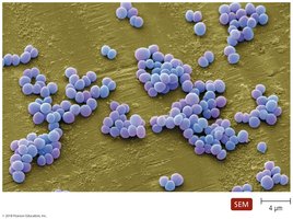
Most commonly caused by Staphylococcus species, which are facultatively anaerobic, Gram-positive cocci arranged in clusters. Two main species:
Staphylococcus epidermidis: Major member of the microbiome, rarely pathogenic.
Staphylococcus aureus: More virulent, produces a variety of disease conditions.
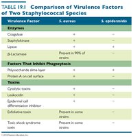
Virulence Factors of Staphylococci
Enzymes (coagulase, staphylokinase, lipase, β-lactamase)
Structures that inhibit phagocytosis (capsule, Protein A)
Toxins (cytolysins, leukocidins, exfoliative toxins, toxic shock syndrome toxin)
Virulence Factor | S. aureus | S. epidermidis |
|---|---|---|
Coagulase | + | - |
Staphylokinase | + | - |
Lipase | + | + |
β-Lactamase | Present in 90% of strains | - |
Polysaccharide slime layer | + | + |
Protein A | + | - |
Cytolysins | + | - |
Leukocidin | + | - |
Exfoliative toxin | Present in some strains | - |
Toxic shock syndrome toxin | Present in some strains | - |
Pathogenesis
Transmitted via direct contact or fomites.
Bacteria invade hair follicles and sebaceous glands, causing inflammation and pus formation.
Can spread to form furuncles, carbuncles, or enter the bloodstream.
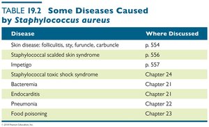
Diseases Caused by Staphylococcus aureus
Disease | Where Discussed |
|---|---|
Skin disease: folliculitis, sty, furuncle, carbuncle | p. 554 |
Staphylococcal scalded skin syndrome | p. 556 |
Impetigo | p. 557 |
Staphylococcal toxic shock syndrome | Chapter 24 |
Bacteremia | Chapter 21 |
Endocarditis | Chapter 21 |
Pneumonia | Chapter 22 |
Food poisoning | Chapter 23 |
Epidemiology
S. epidermidis: Common on skin, rarely causes disease except in immunocompromised or with prosthetic devices.
S. aureus: Not a permanent resident but often found in nostrils and moist skin folds; about 20% of people are asymptomatic carriers.
Diagnosis, Treatment, and Prevention
Diagnosis: Detection of Gram-positive cocci in clusters from pus.
Treatment: Topical mupirocin, dicloxacillin, vancomycin for resistant strains.
Prevention: Aseptic techniques in hospitals.
Staphylococcal Scalded Skin Syndrome (SSSS)
Signs and Symptoms
Red, wrinkled skin that forms blisters and peels off in sheets.
Blisters contain clear fluid without bacteria or white blood cells.
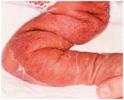
Pathogen and Virulence Factors
Caused by S. aureus strains producing exfoliative toxins.
Toxins cause dissolution of desmosomes, leading to epidermal separation.
Pathogenesis
Toxins spread via blood (toxemia), causing widespread skin damage.
No scarring as the dermis is unaffected; death is rare but can occur due to secondary infections.
Epidemiology, Diagnosis, and Treatment
Primarily affects infants and children under 5.
Diagnosed by characteristic skin sloughing; treated with IV antibiotics (e.g., Nafcillin, oxacillin).
Prevention is difficult due to the ubiquity of S. aureus.
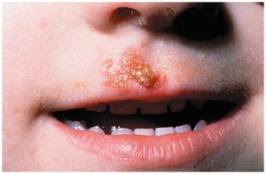
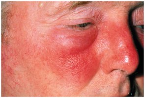
Impetigo (Pyoderma) and Erysipelas
Signs and Symptoms
Impetigo: Red patches on face and limbs, develop into oozing, pus-filled vesicles that form a honey-colored crust.
Erysipelas: Impetigo infection spreads to lymph nodes, causing red areas with distinct margins, swollen nodes, pain, fever, and leukocytosis.
Pathogens and Virulence Factors
Most impetigo cases caused by S. aureus (80%), some by Streptococcus pyogenes (Group A Streptococcus).
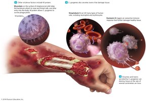
S. pyogenes virulence factors: M protein, hyaluronic acid capsule, pyrogenic toxins.
Pathogenesis and Epidemiology
Bacteria colonize compromised skin; can spread to blood and kidneys (acute glomerulonephritis).
Transmitted by direct contact or fomites; most common in children (2-5 years) and elderly.
Diagnosis, Treatment, and Prevention
Diagnosis: Vesicles of impetigo are diagnostic.
Treatment: Oral/topical antimicrobials for impetigo, penicillin for erysipelas.
Prevention: Proper hygiene and cleanliness.
Necrotizing Fasciitis (“Flesh-Eating Bacteria”)
Signs and Symptoms
Redness, intense pain, swelling, fever, nausea, muscle aches, and possible mental confusion.
Tissue destruction leads to decreased blood pressure and coma.
Pathogen and Virulence Factors
Most cases caused by S. pyogenes (Group A Streptococcus).
Virulence factors: Enzymes and toxins that allow tissue invasion, resist phagocytosis, and damage cells.
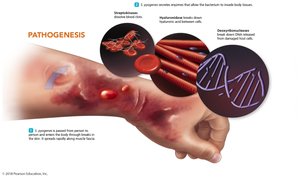
Pathogenesis, Epidemiology, and Treatment
Transmitted via breaks in the skin; spreads rapidly through tissues.
Early diagnosis is difficult; requires surgical removal of dead tissue and antibiotics (clindamycin, penicillin).
Prevention is challenging due to the common presence of S. pyogenes.
Acne
Pathogen
Commonly caused by Propionibacterium acnes, a Gram-positive, rod-shaped diphtheroid.
Also caused by Staphylococcus aureus.
Pathogenesis and Epidemiology
Normal microbiome member; overgrows due to increased sebum during adolescence.
Begins at adolescence but can occur later in life.
Diagnosis, Treatment, and Prevention
Diagnosis: Visual examination.
Treatment: Antimicrobials, exfoliative drugs, Accutane (reduces sebum), ultraviolet light.
Cat Scratch Disease
Signs and Symptoms
Fever, malaise, localized swelling at infection site and lymph nodes.
Pathogen and Virulence Factors
Caused by Bartonella henselae; endotoxin is the main virulence factor.
Pathogenesis and Epidemiology
Transmitted by cat bites/scratches or blood-sucking arthropods (fleas).
Diagnosis, Treatment, and Prevention
Diagnosis: Serological testing.
Treatment: Antimicrobials.
