 Back
BackMicrobial Diseases of the Skin and Eye: Mechanisms, Pathogens, and Host Responses
Study Guide - Smart Notes
 Tailored notes based on your materials, expanded with key definitions, examples, and context.
Microbial Diseases: Overview
Microbial diseases, also known as infectious or communicable diseases, are illnesses caused by microscopic organisms (pathogens) that invade the body. While many microbes are beneficial, such as those in the gut microbiome, pathogenic microbes can disrupt cellular activities or release toxins that cause illness.
Pathogens: Include bacteria, viruses, fungi, and parasites.
Transmission: Can occur via direct contact, contaminated objects, or vectors.
Host Response: The immune system mounts defenses to eliminate pathogens, but some microbes evade or suppress these responses.
Microbial Diseases of the Skin and Eye
Skin and eye infections are often caused by similar pathogens, such as Staphylococcus and Streptococcus, which thrive in warm, moist environments. These infections range from minor irritations to severe, sight-threatening conditions.
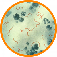
Bacterial Infections of the Skin
Bacterial pathogens typically enter through small cuts, hair follicles, or mucous membranes. The most common causative agent is Staphylococcus aureus.
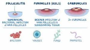
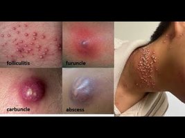
Types of Skin Infections
Folliculitis: Superficial infection of hair follicles, presenting as small, itchy, or tender pus-filled bumps.
Furuncle (Boil): Deeper infection of hair follicles and surrounding tissue, resulting in a painful, necrotic, pus-filled abscess.
Carbuncle: Cluster of interconnected furuncles forming a larger area of infection, often with multiple drainage points and systemic symptoms like fever and chills.
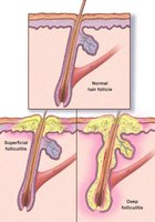
Pathophysiological Process
Initiation: Pathogenic bacteria invade the superficial part of a hair follicle, triggering an inflammatory response with neutrophil migration.
Progression: If not contained, infection extends deeper, causing tissue necrosis and abscess formation (furuncle).
Advanced Stage: Multiple furuncles may coalesce into a carbuncle, with deeper tissue involvement and systemic symptoms.

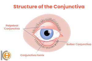
Bacterial Infections of the Eye
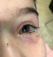
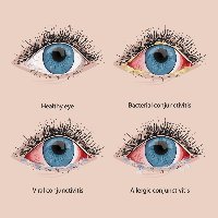
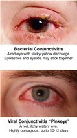
Bacterial conjunctivitis ("pink eye") is an inflammation of the eye's outer membrane, often causing a thick, yellowish discharge. Common pathogens include Staphylococcus aureus, Streptococcus pneumoniae, and Haemophilus influenzae.
Transmission: Direct contact (e.g., fingers), contaminated objects (towels, cosmetics, contact lenses), or spread from adjacent mucosal tissues.
Mechanism: Colonization of the conjunctiva leads to a breach of the epithelial barrier, followed by an acute inflammatory response.
Inflammatory Cascade in Bacterial Conjunctivitis
Vasodilation: Blood vessels in the conjunctiva dilate, causing redness (hyperemia).
Vascular Leakage: Increased permeability leads to swelling (edema or chemosis).
Cellular Exudate: Neutrophils migrate to the area, mixing with dead bacteria and debris to form mucopurulent or purulent discharge ("glued eyes").
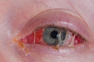
Secondary Defenses: Increased tear production and blinking help rinse bacteria; tears contain lysozyme and immunoglobulins that inhibit bacterial growth.
Viral Infections of the Skin and Eye
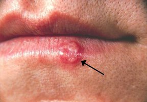
Viruses can cause systemic rashes or localized lesions affecting both the skin and eyes. Herpes Simplex Virus Type 1 (HSV-1) is a major cause of cold sores and can lead to Herpes Keratitis, a significant cause of corneal blindness.
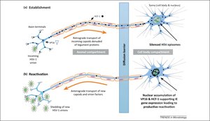
HSV-1 Infection Mechanism
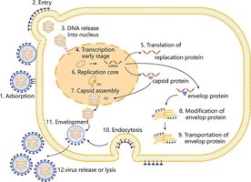
Lytic Phase: Virus infects surface cells, attaches, fuses, and delivers DNA to the nucleus for replication.
Replication: Occurs in three stages—Immediate-Early (IE), Early (E), and Late (L)—to produce new virus particles.
Latency: Virus remains dormant in nerve cells (e.g., trigeminal ganglion), with most genes silenced except for latency-associated transcripts (LAT).
Reactivation: Stress or fever can trigger reactivation, leading to recurrent outbreaks.
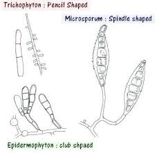
Fungal Infections of the Skin (Cutaneous Mycoses)
Cutaneous mycoses, such as ringworm and athlete's foot, are caused by dermatophytes that feed on keratin in the skin. These infections are usually restricted to the non-living, keratinized layers of the skin.
Adhesion and Growth: Fungal spores adhere to skin cells and germinate in warm, moist conditions.
Keratin Degradation: Dermatophytes secrete chemicals and enzymes to break down keratin for nutrient acquisition.
Immune Response: Inflammation leads to redness, itching, and scaling. Some fungi evade or suppress immune responses, causing chronic infection.
Host Defense: Increased skin turnover and scaling help shed infected cells.
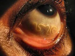
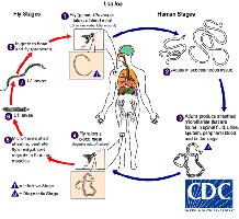
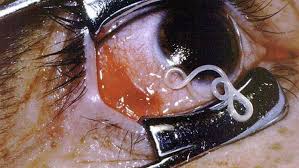
Parasitic Infections of the Eye: Loa loa (African Eye Worm)
Loa loa infection is notable for the visible migration of the adult worm across the eye's surface, though the eye is not the parasite's final destination.
Transmission: Bite from a Chrysops fly deposits larvae, which mature into adult worms under the skin.
Eye Migration: Adult worms may temporarily pass under the conjunctiva, causing irritation and visible movement.
Symptoms: Eye irritation, itching, and localized swelling (Calabar swellings) due to immune reaction. Microfilariae circulate in the blood during the day.
Treatment: Physical removal of the worm and antiparasitic medications; caution is needed to avoid severe inflammation from rapid parasite death.
Sample Exam Questions
A patient presents with a scaling ring-like lesion on the skin. Which of the following events is most critical for initiating nutrient acquisition by dermatophytes? Answer: B. Secretion of sulfite disrupting disulfide bonds in keratin
Which of the following best explains how HSV-1 persists lifelong in neurons without being eliminated? Answer: C. Expression of latency-associated transcripts (LAT) with silencing of lytic genes
The “glued eye” symptom seen in bacterial conjunctivitis is primarily due to: Answer: C. Accumulation of neutrophils, bacteria, and debris forming purulent exudate
