 Back
BackMicrobial Eye Infections
Study Guide - Smart Notes
 Tailored notes based on your materials, expanded with key definitions, examples, and context.
Eye Infections: Structure, Defenses, and Pathogenesis
Basic Structure and Defense Mechanisms of the Eye
The human eye is a complex organ with specialized structures and defense mechanisms that protect against microbial invasion. Understanding these features is essential for comprehending how infections develop and are prevented.
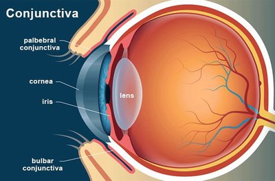
Cornea: The transparent, multi-layered structure at the front of the eye, covering the iris and pupil. It consists of 5–6 layers of epithelial cells that can rapidly regenerate if damaged.
Conjunctiva: A thin epithelial membrane that covers the eyeball and lines the eyelids, surrounding the cornea and providing a barrier to pathogens.
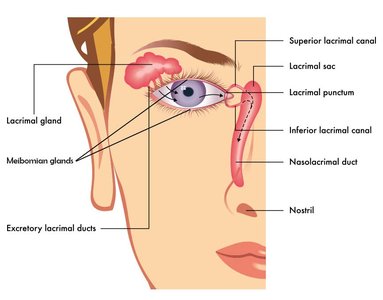
Lacrimal Gland and Tear Film: The lacrimal gland produces tears, which contain water, oils, mucus, lysozyme (an enzyme that breaks down bacterial cell walls), and lactoferrin (which binds free iron, limiting bacterial growth). Tears rinse away debris and microbes.
Eyeball Structure: The thick outer wall of the eyeball prevents microbial penetration.
Ocular Microbiome
The surface of the eye is colonized by a diverse microbiome, which plays a role in maintaining ocular health and preventing infection.
Resident Bacteria: The cornea and conjunctiva are colonized by various bacterial phyla, including Actinobacteria, Firmicutes, and Proteobacteria.
Microbiome Distribution: Different bacterial species dominate the conjunctiva and cornea, as shown by the Ocular Microbiome Project.
Types of Microbial Eye Infections
Eye infections can be caused by bacteria, viruses, fungi, and parasites. The conjunctiva and cornea are the most common targets. Contact lens use increases the risk of infection, especially with poor hygiene.
Conjunctivitis (Pink Eye)
Conjunctivitis is the inflammation of the conjunctiva, presenting as redness, swelling, and discharge. It can be caused by infectious or non-infectious agents.
Non-contagious Conjunctivitis: Triggered by allergens, chemicals, or foreign objects.
Contagious Conjunctivitis: Caused by bacteria or viruses.
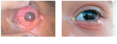
Symptoms: Red or pink sclera, swollen conjunctiva, discharge (watery or purulent), and eyelids that may stick together.
Viral Conjunctivitis
Most commonly caused by adenoviruses, but herpes viruses can also be involved. Highly contagious and often associated with upper respiratory infections.
Symptoms: Begins in one eye, spreads to the other, red/swollen conjunctiva, watery discharge, itching, and pain.
Transmission: Direct contact, fomites, or vertical transmission (e.g., HSV during birth).
Prevention: Hand hygiene, avoiding shared personal items, and not wearing contacts during infection.
Treatment: Usually self-limiting; severe cases may require medical attention.
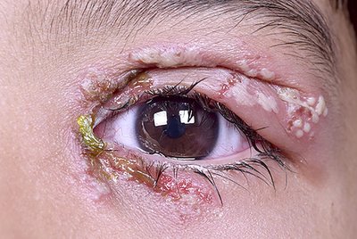
Herpetic Eye Infections: Simplex vs. Zoster
Herpes simplex virus (HSV) and varicella-zoster virus (VZV) can both cause recurrent eye infections, leading to scarring and vision loss.
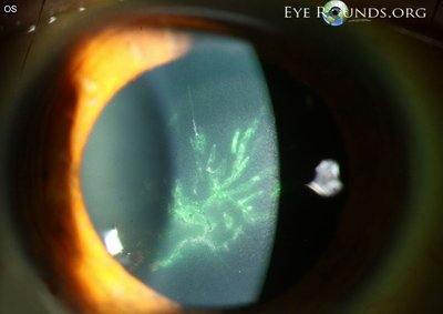
HSV (Simplex): Dendritic lesions with terminal bulbs; treated with topical antivirals.
VZV (Zoster): Pseudodendritic lesions with tapered ends; treated with systemic antivirals and vaccination.


Bacterial Conjunctivitis
Commonly caused by Haemophilus influenzae, Staphylococcus spp., Streptococcus spp., and Moraxella spp. Neonatal cases may involve Neisseria gonorrhoeae or Chlamydia trachomatis.
Symptoms: Similar to viral conjunctivitis, but with thick, pus-like (yellow/green) discharge and crusting.
Neonatal Conjunctivitis: Can cause serious damage; prevented with antibiotic drops at birth.
Treatment: Often self-limiting; antibiotics may be prescribed.
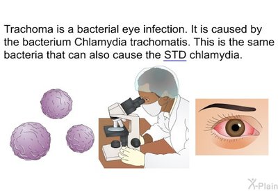
Trachoma
Trachoma is a chronic bacterial infection caused by certain serotypes of Chlamydia trachomatis. It is the leading cause of preventable microbial blindness worldwide.
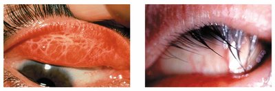
Symptoms: Rough appearance of the conjunctiva, scarring, and inward turning of the eyelid (trichiasis), leading to corneal damage.
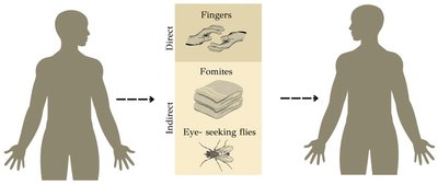
Transmission: Direct contact, fomites, and eye-seeking flies; associated with poor sanitation.
Prevention: Facial cleanliness, improved sanitation, and public health education.
Treatment: Oral azithromycin, topical antibiotics, and surgery for advanced cases.
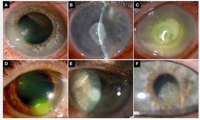
Keratitis
Keratitis is a severe inflammation of the cornea that can lead to vision loss if untreated. It may be caused by viruses, bacteria, fungi, or parasites.
Symptoms: Eye pain, blurred vision, sensitivity to light, and discharge.
Urgency: Keratitis is more serious than conjunctivitis and requires immediate medical attention.
Viral Keratitis
Most often caused by herpes simplex virus type 1 (HSV-1), which reactivates via the ophthalmic nerve.
Symptoms: Conjunctivitis, pain, blurred vision, photophobia, watery discharge.
Bacterial Keratitis
Commonly caused by Pseudomonas aeruginosa and Staphylococcus aureus. Risk factors include improper contact lens use and eye injury.
Symptoms: Similar to viral keratitis, with additional discharge and tearing.
Prevention: Proper contact lens hygiene and hand washing.
Treatment: Antibiotic eye drops.
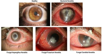
Fungal Keratitis
Caused by fungi such as Fusarium, Aspergillus, and Candida species. Risk factors include eye trauma, surgery, immunosuppression, and contact lens use.
Symptoms: Similar to bacterial keratitis, but often more difficult to treat.
Treatment: Prolonged antifungal therapy; surgery may be necessary.
Parasitic Keratitis
Most commonly caused by Acanthamoeba species (protozoa), found in water and associated with improper contact lens care. Helminthic keratitis can be caused by Onchocerca volvulus (river blindness).
Acanthamoeba Keratitis: Rare but serious; not contagious. Early diagnosis and treatment are critical.
River Blindness (Ocular Onchocerciasis): Transmitted by blackfly bites; larvae migrate to the eye, causing inflammation and blindness.
Treatment: Antiparasitic drugs (e.g., ivermectin, doxycycline for river blindness).
Summary Table: Eye Infections
The following table summarizes the main types of microbial eye infections, their causes, transmission, and key features.
Infection | Etiological Agent | Transmission | Key Features |
|---|---|---|---|
Conjunctivitis (Viral) | Adenoviruses, HSV | Respiratory droplets, direct contact, fomites | Watery discharge, red eye, highly contagious |
Conjunctivitis (Bacterial) | H. influenzae, Staphylococcus, Streptococcus, Moraxella | Direct contact, fomites | Pus-like discharge, eyelids stuck, more severe in neonates |
Trachoma | Chlamydia trachomatis | Fomites, fingers, flies | Scarring, trichiasis, leading cause of infectious blindness |
Keratitis (Viral) | HSV-1 | Reactivation, direct contact | Dendritic corneal lesions, pain, vision loss |
Keratitis (Bacterial) | P. aeruginosa, S. aureus | Contact lens use, injury | Corneal ulcer, discharge, rapid progression |
Keratitis (Fungal) | Fusarium, Aspergillus, Candida | Trauma, surgery, contact lens | Chronic, difficult to treat, may require surgery |
Keratitis (Parasitic) | Acanthamoeba, Onchocerca volvulus | Contaminated water, blackfly bite | Severe pain, vision loss, river blindness |
