 Back
BackMicrobial Genetics: Foundations, Experiments, and Molecular Mechanisms
Study Guide - Smart Notes
 Tailored notes based on your materials, expanded with key definitions, examples, and context.
Microbial Genetics: Foundations, Experiments, and Molecular Mechanisms
Introduction to Microbial Genetics
Microbial genetics is the study of the mechanisms of heritable information in microorganisms, focusing on the structure, function, and regulation of genetic material. This field underpins our understanding of microbial physiology, evolution, and biotechnology.
Historical Experiments Establishing DNA as Genetic Material
The Griffith Experiment: Discovery of Transformation
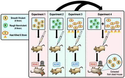
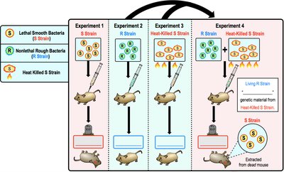
In 1928, Frederick Griffith demonstrated that bacteria can transfer genetic material through a process called transformation. He used two strains of Streptococcus pneumoniae: a smooth, virulent (S) strain and a rough, nonvirulent (R) strain. When mice were injected with a mixture of heat-killed S strain and live R strain, the mice died, and live S strain bacteria could be recovered, indicating that genetic material from the dead S strain transformed the R strain into a virulent form.
Transformation: The uptake and incorporation of external DNA by a cell, resulting in genetic and phenotypic change.
Key Finding: Genetic information can be transferred horizontally between bacteria.
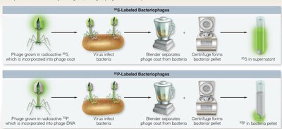
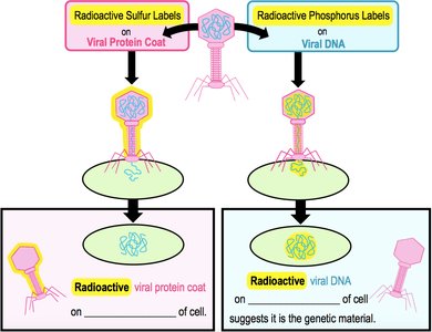
The Hershey-Chase Experiment: DNA is the Genetic Material
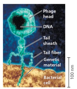
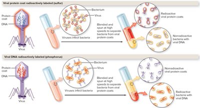
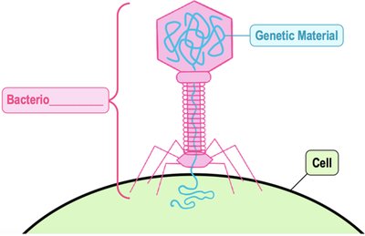
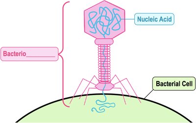
In 1952, Martha Hershey and Alfred Chase used bacteriophages (viruses that infect bacteria) to confirm that DNA, not protein, is the genetic material. They labeled phage protein coats with radioactive sulfur (35S) and DNA with radioactive phosphorus (32P). Only the radioactive DNA entered the bacterial cells and directed viral replication, proving that DNA carries genetic information.
Bacteriophage: A virus that infects bacteria, consisting of a protein coat and nucleic acid core.
Key Finding: Only DNA, not protein, is inherited during viral infection of bacteria.
Chargaff’s Rules and DNA Composition
Chargaff’s Rules
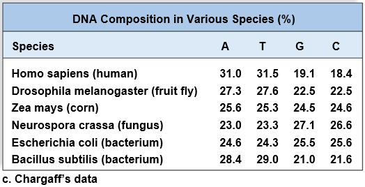
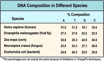
Erwin Chargaff discovered that the composition of DNA varies between species, but within each species, the amount of adenine (A) is approximately equal to thymine (T), and the amount of guanine (G) is approximately equal to cytosine (C). This provided key evidence for base pairing in DNA structure.
Key Rule: %A ≈ %T and %G ≈ %C in DNA of any given species.
Implication: Base pairing is fundamental to DNA structure and replication.
Species | A | T | G | C |
|---|---|---|---|---|
Homo sapiens (human) | 31.0 | 31.5 | 19.1 | 18.4 |
Drosophila melanogaster (fruit fly) | 27.3 | 27.6 | 22.5 | 22.5 |
Zea mays (corn) | 25.6 | 25.3 | 24.5 | 24.6 |
Neurospora crassa (fungus) | 23.0 | 23.3 | 27.1 | 26.6 |
Escherichia coli (bacterium) | 24.6 | 24.3 | 25.5 | 25.6 |
Bacillus subtilis (bacterium) | 28.4 | 29.0 | 21.0 | 21.6 |
Discovery of DNA Structure
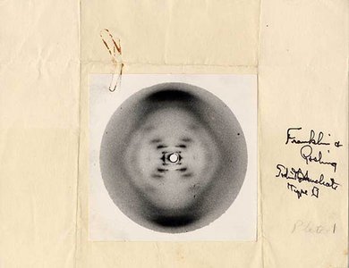
X-ray Diffraction and the Double Helix
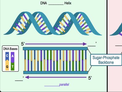
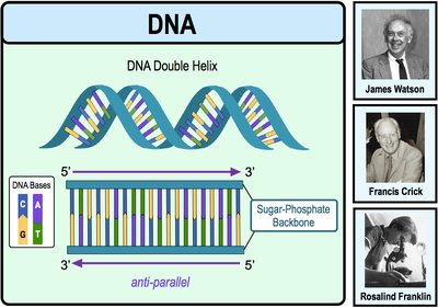
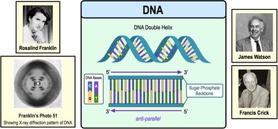
Rosalind Franklin used X-ray diffraction to capture images of DNA, notably Photo 51, which revealed the helical structure of DNA. James Watson and Francis Crick used these data to propose the double helix model in 1953, describing DNA as two antiparallel strands held together by specific base pairing (A–T, C–G) via hydrogen bonds.
Double Helix: Two antiparallel strands of nucleotides twisted into a helical shape.
Base Pairing: Adenine pairs with thymine (A–T), and guanine pairs with cytosine (G–C).
DNA Replication: Mechanisms and Enzymes
Phosphodiester Bond Formation
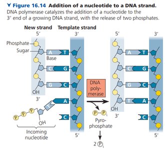
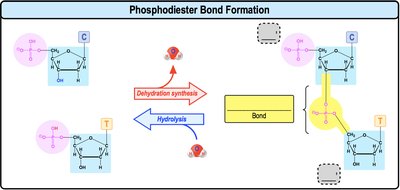
During DNA replication, nucleotides are joined by phosphodiester bonds between the 3' hydroxyl group of one nucleotide and the 5' phosphate group of the next. This reaction is catalyzed by DNA polymerase and is essential for the elongation of the DNA strand.
Directionality: DNA is synthesized in the 5' to 3' direction.
Enzyme: DNA polymerase catalyzes the addition of nucleotides.
Additional info: The above sections cover the foundational experiments and molecular mechanisms that established DNA as the genetic material and revealed its structure and replication. These concepts are essential for understanding microbial genetics and the molecular biology of all living organisms.
